 Indietro
Chapter 12: Nervous Tissue – Structure, Function, and Physiology
Guida di studio - Note intelligenti
 Appunti personalizzati basati sui tuoi materiali, ampliati con definizioni chiave, esempi e contesto.
Nervous Tissue: Structure and Function
Anatomical and Functional Divisions of the Nervous System
The nervous system is divided into central and peripheral components, each with specialized functions for processing and transmitting information throughout the body.
Central Nervous System (CNS): Consists of the brain and spinal cord; responsible for integrating, processing, and coordinating sensory input and motor output.
Peripheral Nervous System (PNS): Composed of nervous tissue outside the CNS; includes sensory and motor divisions.
Afferent Division: Carries sensory information from receptors to the CNS.
Efferent Division: Transmits motor commands from the CNS to effectors (muscles and glands).
Somatic Nervous System (SNS): Controls voluntary movements of skeletal muscles.
Autonomic Nervous System (ANS): Regulates involuntary functions; subdivided into sympathetic and parasympathetic divisions.
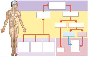
Additional info: The ENS (enteric nervous system) is sometimes considered a third division, controlling gastrointestinal function.
Structure of a Typical Neuron
Neurons are the functional units of the nervous system, specialized for transmitting electrical impulses.
Dendrites: Receive incoming signals from other neurons.
Cell Body (Soma): Contains the nucleus and organelles; integrates signals.
Axon Hillock: Region where action potentials are initiated.
Axon: Conducts action potentials away from the cell body.
Axon Terminals: Release neurotransmitters to communicate with other cells.
Nissl Bodies: Rough endoplasmic reticulum and ribosomes; responsible for protein synthesis.
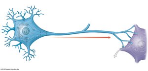
Example: Motor neurons transmit signals from the CNS to muscles, causing contraction.
Classification of Neurons
Neurons are classified based on their structure and function.
Structural Classification:
Anaxonic: No obvious axon; found in CNS.
Bipolar: Two processes; found in retina, olfactory epithelium.
Unipolar: Single elongated process; sensory neurons in PNS.
Multipolar: One axon, multiple dendrites; most common in CNS.
Functional Classification:
Sensory (Afferent) Neurons: Carry information to CNS.
Motor (Efferent) Neurons: Carry commands from CNS to effectors.
Interneurons: Integrate information and coordinate responses.
Example: Bipolar neurons are essential for vision and smell.
Neuroglia: Support Cells of the Nervous System
Neuroglia are non-neuronal cells that provide structural and functional support to neurons.
CNS Neuroglia:
Astrocytes: Maintain blood-brain barrier, regulate ion/nutrient levels, form scar tissue.
Ependymal Cells: Line ventricles and central canal; produce and circulate cerebrospinal fluid.
Oligodendrocytes: Myelinate CNS axons; provide structural framework.
Microglia: Phagocytose debris and pathogens.
PNS Neuroglia:
Satellite Cells: Surround neuron cell bodies in ganglia; regulate environment.
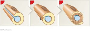
Schwann Cells: Myelinate PNS axons; assist in repair after injury.
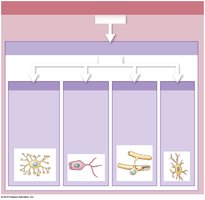
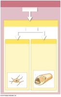
Example: Oligodendrocytes are responsible for myelination in the CNS, while Schwann cells perform this function in the PNS.
Myelination and Nerve Regeneration
Myelination increases the speed of nerve impulse conduction and is essential for normal nervous system function.
Schwann Cells: Wrap around axons in the PNS, forming the myelin sheath.
Nodes of Ranvier: Gaps in the myelin sheath where action potentials are regenerated.
Neurilemma: Outermost layer of Schwann cell cytoplasm; aids in regeneration.
Nerve Regeneration: Schwann cells guide regrowth in PNS; limited regeneration in CNS due to astrocyte scar formation.
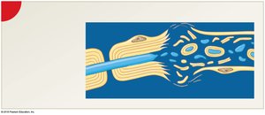
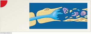
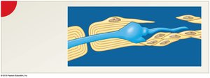
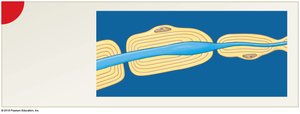
Example: Peripheral nerves can regenerate after injury if the neurilemma remains intact.
Resting Membrane Potential
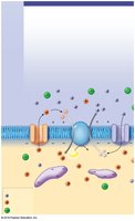
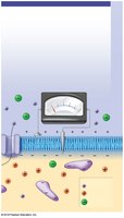
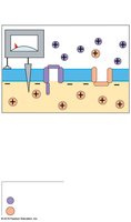
The resting membrane potential is the electrical charge difference across the plasma membrane of a neuron at rest.
Passive Chemical Gradients: K+ moves out, Na+ moves in, driven by concentration gradients.
Active Na+/K+ Pump: Maintains gradients by pumping 3 Na+ out and 2 K+ in, using ATP.
Electrical Gradient: More positive charges outside, more negative inside due to impermeable proteins.
Typical Resting Potential: About −70 mV.
Equation:
Example: The Na+/K+ ATPase is essential for maintaining neuronal excitability.
Gated Ion Channels and Graded Potentials
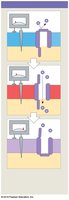
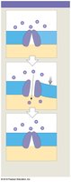
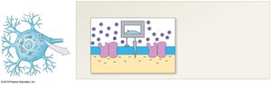
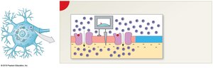
Neuronal membranes contain gated ion channels that open or close in response to specific stimuli, leading to changes in membrane potential.
Chemically Gated Channels: Open in response to neurotransmitter binding (e.g., ACh).
Voltage-Gated Channels: Open in response to changes in membrane potential.
Mechanically Gated Channels: Open in response to physical distortion.
Graded Potentials: Local changes in membrane potential; can lead to action potentials if threshold is reached.
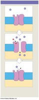
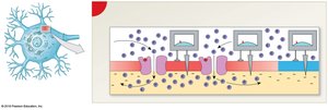
Example: Chemically gated channels are crucial for synaptic transmission.
Depolarization, Repolarization, and Hyperpolarization
Changes in membrane potential are essential for neuronal signaling.
Depolarization: Membrane potential becomes less negative (closer to zero).
Repolarization: Return to resting membrane potential after depolarization.
Hyperpolarization: Membrane potential becomes more negative than resting.
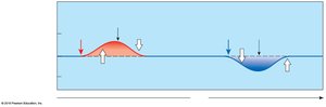
Example: Inhibitory neurotransmitters cause hyperpolarization, reducing neuronal excitability.
Generation and Propagation of Action Potentials
Action potentials are rapid, all-or-none electrical signals that travel along axons.
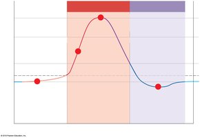
Threshold: Minimum depolarization required to trigger an action potential (typically −60 mV).
All-or-None Principle: Action potentials are always the same size; either triggered or not.
Phases:
Depolarization: Voltage-gated Na+ channels open; Na+ influx.
Repolarization: Na+ channels inactivate; K+ channels open; K+ efflux.
Hyperpolarization: K+ channels remain open briefly.
Restoration: Na+/K+ pump restores resting potential.
Refractory Periods:
Absolute: No new action potential can be generated.
Relative: Only a stronger stimulus can trigger an action potential.
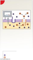
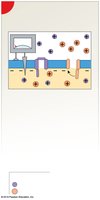
Equation: (Ohm's law for ion flow)
Example: Action potentials are essential for communication between neurons and muscle cells.
Propagation of Action Potentials
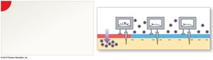
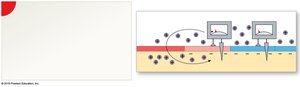
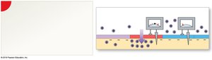
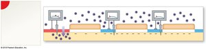
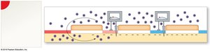
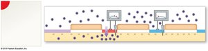
Action potentials propagate along axons by two mechanisms: continuous and saltatory conduction.
Continuous Propagation: Occurs in unmyelinated axons; action potential moves stepwise along the entire membrane.
Saltatory Propagation: Occurs in myelinated axons; action potential jumps from node to node, increasing speed.
Fiber Types:
Type A: Large, myelinated, fast (120 m/sec).
Type B: Medium, myelinated, intermediate speed (18 m/sec).
Type C: Small, unmyelinated, slow (1 m/sec).
Example: Type A fibers are used for rapid motor responses, such as reflexes.
Structure and Function of Synapses
Synapses are specialized junctions where neurons communicate with other cells.
Electrical Synapses: Direct physical contact via gap junctions; rapid transmission.
Chemical Synapses: Neurotransmitters cross a synaptic cleft; more common; involve synaptic delay.
Components: Presynaptic membrane, synaptic cleft, postsynaptic membrane.
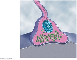
Example: Neuromuscular junctions are chemical synapses between motor neurons and muscle fibers.
Events at a Cholinergic Synapse
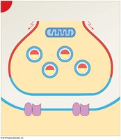
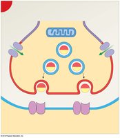
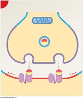
Cholinergic synapses use acetylcholine (ACh) as their neurotransmitter.
Step 1: Action potential depolarizes axon terminal.
Step 2: Voltage-gated Ca2+ channels open; Ca2+ influx triggers ACh release.
Step 3: ACh binds to postsynaptic receptors; Na+ channels open; graded depolarization occurs.
Step 4: ACh is broken down by acetylcholinesterase (AChE); choline is reabsorbed and recycled.
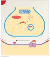
Example: Cholinergic synapses are critical for muscle contraction and memory.
Neurotransmitters and Neuromodulators
Neurotransmitters are chemicals that transmit signals across synapses; neuromodulators modify synaptic activity.
Excitatory Neurotransmitters: Cause depolarization; promote action potentials (e.g., ACh, dopamine, glutamate, serotonin).
Inhibitory Neurotransmitters: Cause hyperpolarization; suppress action potentials (e.g., GABA, dopamine, serotonin).
xasqwk jhtyr432ert5yk ,jyNeurotransmitter | Function | Examples of Malfunctions |
|---|---|---|
Acetylcholine (ACh) | Enables muscle action, learning, memory | Alzheimer's disease: ACh-producing neurons deteriorate |
Dopamine | Influences movement, learning, attention, emotion | Schizophrenia (oversupply), Parkinson's (undersupply) |
Serotonin | Affects mood, hunger, sleep, arousal | Depression (undersupply) |
Norepinephrine | Controls alertness, arousal | Depressed mood (undersupply) |
GABA | Major inhibitory neurotransmitter | Seizures, tremors, insomnia (undersupply) |
Glutamate | Major excitatory neurotransmitter | Overstimulation: migraines, seizures |
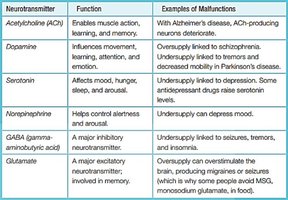
Example: Glutamate is involved in learning and memory; GABA inhibits excessive neuronal activity.
Mechanisms of Neurotransmitter Action
Neurotransmitters can act directly or indirectly on postsynaptic cells.
Direct Effects: Bind to ion channels, causing immediate changes in membrane potential (e.g., ACh).
Indirect Effects via G Proteins: Activate second messenger systems (e.g., cAMP) to modulate cell activity.
Indirect Effects via Intracellular Enzymes: Neuromodulators like nitric oxide and carbon monoxide produce long-term changes.
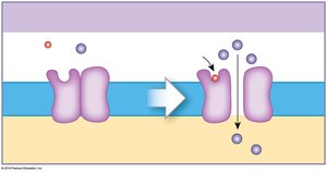
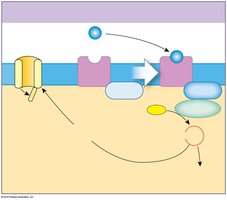
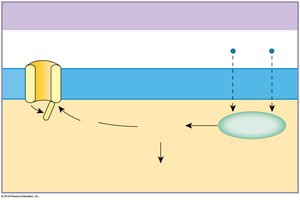
Example: Dopamine can act both directly and indirectly, affecting mood and movement.
Information Processing in Nervous Tissue
Neurons integrate multiple inputs to determine their output, using summation and modulation mechanisms.
Temporal Summation: Multiple stimuli from the same source in rapid succession add together.
Spatial Summation: Simultaneous stimuli from different locations combine their effects.
Excitatory Post-Synaptic Potential (EPSP): Depolarizing effect; increases likelihood of action potential.
Inhibitory Post-Synaptic Potential (IPSP): Hyperpolarizing effect; decreases likelihood of action potential.
Presynaptic Inhibition: Reduces neurotransmitter release (e.g., GABA).
Presynaptic Facilitation: Increases neurotransmitter release (e.g., serotonin).
Example: Summation allows neurons to integrate complex information and make decisions about firing.
Additional info: These mechanisms are fundamental for higher-order functions such as memory, learning, and planning.
