 Indietro
Chapter 12: The Central Nervous System – Structure and Function
Guida di studio - Note intelligenti
 Appunti personalizzati basati sui tuoi materiali, ampliati con definizioni chiave, esempi e contesto.
The Central Nervous System (CNS)
Overview and Organization
The central nervous system (CNS) consists of the brain and spinal cord, serving as the main control center for the body. It is responsible for integrating sensory information, coordinating motor output, and higher mental functions.
Cephalization: Evolutionary development of the anterior CNS, resulting in increased neuron number and complexity, highest in humans.
Major Regions: Adult brains have four regions:
Cerebral hemispheres
Diencephalon
Brain stem (midbrain, pons, medulla)
Cerebellum
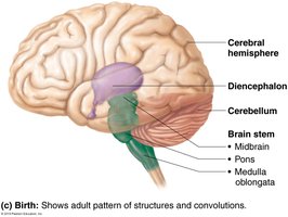
Gray Matter and White Matter
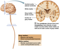
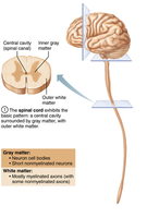
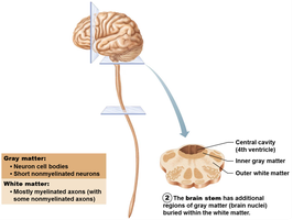
The CNS is organized with a central cavity surrounded by gray matter, and white matter external to gray matter.
Gray Matter: Contains neuron cell bodies and short, nonmyelinated neurons.
White Matter: Composed of myelinated and nonmyelinated axons.
Pattern:
Spinal cord: central cavity, inner gray matter, outer white matter.
Brain stem: additional gray matter nuclei within white matter.
Cerebrum and cerebellum: outer cortex of gray matter, scattered gray matter nuclei amid white matter.
Brain Ventricles
Structure and Function
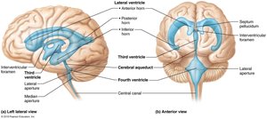
Ventricles are fluid-filled chambers within the brain, continuous with the central canal of the spinal cord.
Cerebrospinal Fluid (CSF): Fills ventricles, provides buoyancy, protection, and nourishment.
Ventricular System:
Paired lateral ventricles (deep in hemispheres, separated by septum pellucidum)
Third ventricle (in diencephalon, connected to lateral ventricles via interventricular foramen)
Fourth ventricle (in hindbrain, connected to third ventricle via cerebral aqueduct, continuous with central canal)
Openings connect fourth ventricle to subarachnoid space: paired lateral apertures and median aperture
Cerebral Hemispheres
Surface Markings and Lobes
The cerebral hemispheres form the superior part of the brain and account for 83% of its mass.
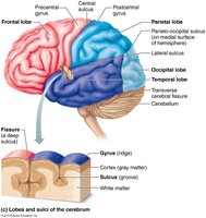
Gyri: Ridges
Sulci: Shallow grooves
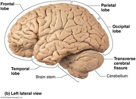
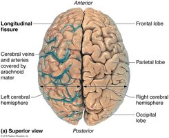
Fissures: Deep grooves (e.g., longitudinal fissure separates hemispheres, transverse fissure separates cerebrum and cerebellum)
Lobes: Frontal, parietal, temporal, occipital, and insula (buried under other lobes)
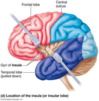
Major Sulci and Regions
Central sulcus: Separates precentral gyrus (frontal lobe) and postcentral gyrus (parietal lobe)
Parieto-occipital sulcus: Separates occipital and parietal lobes
Lateral sulcus: Outlines temporal lobes
Three basic regions:
Cerebral cortex (gray matter, superficial)
White matter (internal)
Basal nuclei (deep within white matter)
Cerebral Cortex
Structure and Function
The cerebral cortex is the "executive suite" of the brain, responsible for conscious mind functions such as awareness, sensory perception, voluntary motor initiation, communication, memory storage, and understanding.
Composition: Thin (2–4 mm) layer of gray matter, 40% of brain mass
Functional Areas:
Motor areas: control voluntary movement
Sensory areas: conscious awareness of sensation
Association areas: integrate diverse information
Contralateral Control: Each hemisphere controls the opposite side of the body
Lateralization: Specialization of function in one hemisphere
Conscious Behavior: Involves the entire cortex
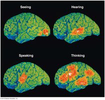
Motor Areas
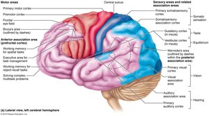
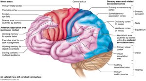
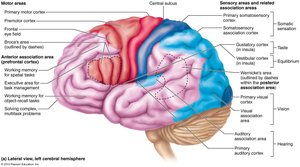
Motor areas are located in the frontal lobe and control voluntary movement.
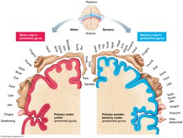
Primary motor cortex: Precentral gyrus; pyramidal cells allow conscious control of precise, skilled movements
Premotor cortex: Plans movements, controls learned and patterned motor skills
Broca’s area: Motor speech area, usually in the left hemisphere
Frontal eye field: Controls voluntary eye movements
Motor Homunculus
Somatotopy maps all muscles of the body to areas on the primary motor cortex. Motor homunculi are caricatures representing contralateral motor innervation. 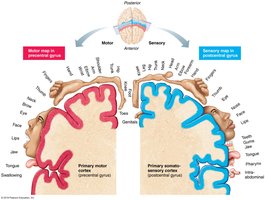
Clinical Note: Motor Cortex Damage
Damage to primary motor cortex (e.g., stroke) causes paralysis on the opposite side of the body.
Muscle strength is not impaired, but voluntary control is lost.
Neuroplasticity allows other neurons to be reprogrammed with practice.
Sensory Areas
Sensory areas are located in parietal, insular, temporal, and occipital lobes.
Primary somatosensory cortex: Postcentral gyri; receives sensory information from skin and proprioceptors
Somatosensory association cortex: Integrates sensory input for object understanding
Visual areas: Primary visual cortex (occipital lobe), visual association area interprets visual stimuli
Auditory areas: Primary auditory cortex (temporal lobe), auditory association area stores sound memories
Vestibular cortex: Awareness of balance
Olfactory cortex: Perception of odors
Gustatory cortex: Perception of taste
Visceral sensory area: Perception of visceral sensations

Multimodal Association Areas
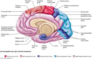
These areas receive inputs from multiple sensory areas and send outputs to multiple areas, allowing integration of sensations, thoughts, and emotions.
Anterior association area (prefrontal cortex): Intellect, cognition, recall, personality
Posterior association area: Pattern and face recognition, spatial awareness, language understanding
Limbic association area: Emotional impact and memory formation
Lateralization of Cortical Function
Lateralization: Division of labor between hemispheres
Cerebral dominance: Hemisphere dominant for language (usually left)
Left hemisphere: Language, math, logic
Right hemisphere: Visual-spatial skills, intuition, emotion, artistic abilities
Cerebral White Matter
Structure and Function
White matter is responsible for communication between cerebral areas and between cortex and lower CNS.
Myelinated fibers: Bundled into large tracts
Fiber Types:
Association fibers: connect different parts of same hemisphere
Commissural fibers: connect gray matter of two hemispheres
Projection fibers: connect hemispheres with lower brain or spinal cord
Internal capsule: Compact band of projection fibers
Corona radiata: Projection fibers radiating through white matter to cortex
Basal Nuclei (Ganglia)
Structure and Function
Basal nuclei are deep within the cerebral hemispheres and play a role in motor control and cognition.
Components: Caudate nucleus, putamen, globus pallidus (caudate + putamen = striatum)
Functions:
Influence muscle movements
Regulate intensity of movements
Filter out incorrect responses
Inhibit unnecessary movements
Disorders: Parkinson’s disease, Huntington’s disease
The Diencephalon
Structure and Components
The diencephalon consists of three paired gray-matter structures: thalamus, hypothalamus, and epithalamus, all enclosing the third ventricle.
Thalamus: Relay station for information to cortex; sorts, edits, and relays ascending input
Epithalamus: Contains pineal gland, secretes melatonin for sleep-wake regulation
Hypothalamus: Main visceral control center; regulates autonomic functions, emotions, body temperature, hunger, thirst, sleep-wake cycles, and endocrine system
Brain Stem
Structure and Function
The brain stem consists of the midbrain, pons, and medulla oblongata. It controls automatic behaviors necessary for survival and connects higher and lower neural centers.
Midbrain: Contains motor tracts, pain suppression nuclei, visual and auditory reflex centers
Pons: Conduction tracts, origin of cranial nerves V-VII, helps regulate breathing
Medulla oblongata: Autonomic reflex center, cardiovascular and respiratory centers, decussation of pyramids (contralateral control)
Cerebellum
Structure and Function
The cerebellum is located dorsal to the pons and medulla, processing input to provide precise, coordinated movements and balance.
Hemispheres: Connected by vermis, each with three lobes
Arbor vitae: Treelike pattern of white matter
Cerebellar processing: Fine-tunes motor activity, receives input from cortex and sensory pathways
Cognitive functions: Role in thinking, language, and emotion
Functional Brain Systems
Limbic System and Reticular Formation
Limbic system: Emotional brain, includes amygdaloid body, cingulate gyrus, hippocampus; involved in memory and emotional responses
Reticular formation: Reticular activating system (RAS) keeps cortex alert, filters stimuli, regulates visceral motor functions
Protection of the Brain
Meninges
Meninges are three connective tissue layers that protect the CNS, blood vessels, and contain CSF.
Pia mater: Delicate, clings to brain, contains blood vessels
Arachnoid mater: Middle layer, spiderweb-like extensions, contains CSF
Dura mater: Strongest, two layers, forms partitions (dural septa) to limit movement
Cerebrospinal Fluid (CSF)
CSF forms a liquid cushion around the brain, providing buoyancy, protection, and nourishment.
Produced by: Choroid plexus
Composition: Similar to plasma, but with less protein and different ion concentrations
Volume: ~150 ml, replaced every 8 hours
Blood Brain Barrier
The blood brain barrier maintains a stable environment for the brain, preventing harmful substances from entering.
Structure: Tight junctions between endothelial cells, astrocyte feet, thick basement membrane
Transport: Simple diffusion, facilitated diffusion, transcytosis
Absent in: Vomiting center, hypothalamus (for monitoring blood)
Spinal Cord
Gross Anatomy and Protection
The spinal cord is enclosed in the vertebral column, beginning at the foramen magnum and ending at L1 or L2.
Functions: Two-way communication, major reflex center
Protection: Bone, meninges, CSF
Spinal dura mater: One layer, does not attach to vertebrae
Epidural space: Fat and veins between vertebrae and dura mater
Site of lumbar puncture: Between L1/L2 and sacrum
Spinal Cord Structure
Conus medullaris: Terminal cone-shaped structure
Filum terminale: Anchors spinal cord to coccyx
Denticulate ligaments: Secure cord to dura mater
Cervical and lumbar enlargements: Nerves serving limbs arise here
Cauda equina: Collection of nerve roots at inferior end
Spinal Cord Cross-sectional Anatomy
Gray matter: Core, butterfly or H-shaped, contains dorsal, ventral, and lateral horns
White matter: Outside, divided into dorsal, lateral, and ventral columns (funiculi)
Central canal: Runs length of cord, filled with CSF
Spinal nerves: 31 pairs, attach by dorsal and ventral roots
Gray matter groups: Somatic sensory, visceral sensory, visceral motor, somatic motor
White matter tracts: Ascending (sensory), descending (motor), transverse (commissural)
Spinal Cord Trauma and Disorders
Transection: Total motor and sensory loss below injury
Paraplegia: Transection between T1 and L1
Quadriplegia: Transection in cervical region
Spinal shock: Temporary loss of function below lesion
Paresthesias: Sensory function loss (dorsal roots/tracts damage)
Paralysis: Motor function loss (ventral roots/horn cells damage)
Flaccid paralysis: No voluntary/involuntary control, muscle atrophy
Spastic paralysis: No voluntary control, muscles stimulated by reflex activity
