 Indietro
Chap 12 CNS - Part 1 - McNeese, Duck
Guida di studio - Note intelligenti
 Appunti personalizzati basati sui tuoi materiali, ampliati con definizioni chiave, esempi e contesto.
Embryonic Development of the Human Brain
Primary and Secondary Brain Vesicles
The human brain develops from the neural tube, which forms three primary brain vesicles: the prosencephalon (forebrain), mesencephalon (midbrain), and rhombencephalon (hindbrain). These vesicles further differentiate into secondary vesicles, giving rise to distinct adult brain structures. - Prosencephalon divides into the telencephalon (cerebral hemispheres) and diencephalon (thalamus, hypothalamus, epithalamus, retina). - Mesencephalon remains as the midbrain. - Rhombencephalon divides into the metencephalon (pons and cerebellum) and myelencephalon (medulla oblongata). - The neural canal forms the ventricular system of the brain and the central canal of the spinal cord. 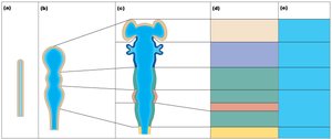
Major Landmarks of the Adult Brain
The adult brain consists of four main regions: cerebrum, diencephalon, brainstem, and cerebellum. - Cerebrum: Largest region, responsible for higher cognitive functions. - Diencephalon: Contains thalamus, hypothalamus, and epithalamus. - Brainstem: Includes midbrain, pons, and medulla oblongata. - Cerebellum: Coordinates movement and balance. 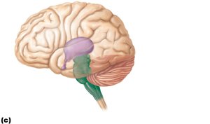
Nervous Tissue and Myelination
Structure of a Neuron
Neurons are the functional units of the nervous system, specialized for transmitting electrical signals. - Dendrites: Receive incoming signals. - Neurosoma: Cell body containing the nucleus. - Axon: Conducts impulses away from the cell body. - Myelin sheath: Insulating layer produced by oligodendrocytes (CNS) or Schwann cells (PNS), increases signal speed. - Node of Ranvier: Gaps in myelin sheath, facilitate saltatory conduction. 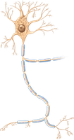
Gray Matter vs. White Matter
Nervous tissue is organized into gray and white matter. - Gray matter: Contains neuron cell bodies and short, nonmyelinated neurons. - White matter: Composed mostly of myelinated axons, responsible for communication between different brain regions.
Distribution in CNS
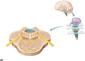
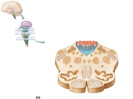
- In the spinal cord: central cavity surrounded by gray matter, with outer white matter. 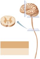 - In the brainstem: additional regions of gray matter (nuclei) are buried within white matter.
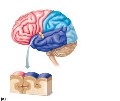
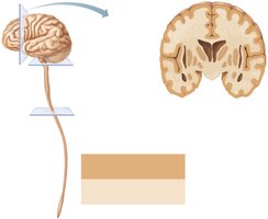 - In the cerebrum and cerebellum: islands of gray matter (nuclei) within white matter, plus an outer cortex of gray matter.
Anatomy of the Cerebrum
Lobes, Sulci, and Fissures
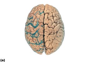
The cerebrum is divided into lobes by prominent sulci and fissures. - Frontal lobe: Associated with motor function, problem solving, and speech. - Parietal lobe: Processes sensory information. - Occipital lobe: Responsible for vision. - Temporal lobe: Involved in hearing and memory. - Insula: Deep within the lateral sulcus, involved in taste and visceral sensation. - Gyri: Ridges of the cortex. - Sulci: Shallow grooves. - Fissures: Deep grooves separating major brain regions. 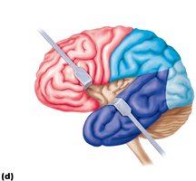
Three Basic Regions of the Hemisphere
- Cerebral cortex: Outer layer of gray matter, site of conscious mind. - White matter: Internal communication pathways. - Basal nuclei: Deep gray matter, involved in motor control and cognition.
Types of White Matter Fibers
- Association fibers: Connect different parts of the same hemisphere. - Commissural fibers: Connect corresponding gray areas of the two hemispheres (e.g., corpus callosum). - Projection fibers: Connect the cortex with lower brain or spinal cord regions. 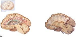
Functional Areas of the Cerebral Cortex
Motor Areas
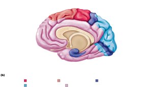
Motor areas are located in the frontal lobe and control voluntary movements. - Primary motor cortex: Located in the precentral gyrus, initiates voluntary movement. - Premotor cortex: Plans movements. - Broca’s area: Controls speech production. - Frontal eye field: Controls voluntary eye movements. 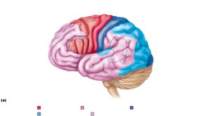
Somatotopy of Functional Areas
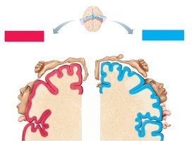
The primary motor and somatosensory cortices are organized as body maps (homunculi), with specific regions controlling or sensing different body parts. 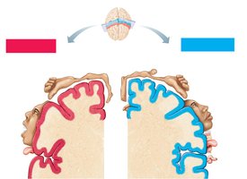
Sensory Areas
Sensory areas are distributed across the parietal, insular, temporal, and occipital lobes. - General senses: Pain, temperature, touch, pressure, vibration, proprioception. - Special senses: Vision, hearing, taste, smell, equilibrium. - Primary somatosensory cortex: Located in the postcentral gyrus, receives sensory input. - Somatosensory association cortex: Integrates sensory information. - Visceral sensory area: Deep in the insula, processes visceral sensations. - Visual, auditory, olfactory, gustatory, vestibular cortices: Process respective special senses. 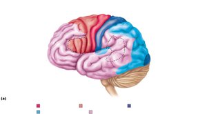
Multimodal Association Areas
These areas integrate information from multiple sensory modalities and are involved in higher cognitive functions. - Anterior association area (prefrontal cortex): Intellect, cognition, recall, personality. - Posterior association area: Recognizing patterns and faces, understanding language (Wernicke’s area). - Limbic association area: Emotional impact and memory formation. 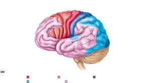
Cerebral Lateralization
The two hemispheres of the cerebrum specialize in different functions. - Left hemisphere: Language, math, logic. - Right hemisphere: Visual-spatial skills, intuition, emotion, artistic and musical abilities. - Cerebral dominance: Refers to the hemisphere dominant for language; 90% of humans are left-dominant.
The Diencephalon
Thalamus
The thalamus is a bilateral structure acting as a relay station for sensory information entering the cortex. - Relay station: All sensory input (except olfaction) passes through the thalamus. - 80% of diencephalon: Largest component. 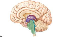
Epithalamus
- Pineal gland: Secretes melatonin, regulates sleep-wake cycles. 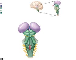
Hypothalamus
The hypothalamus is a major homeostatic control center. - Mammillary bodies: Olfactory relay station. - Infundibulum: Connects hypothalamus to pituitary gland. - Homeostatic functions: Controls autonomic nervous system, emotional responses, body temperature, hunger, thirst, endocrine function, sleep-wake cycles. 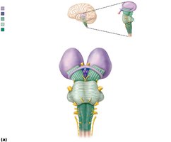
The Brainstem
Structure and Components
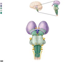
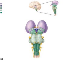
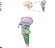
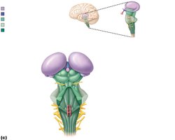
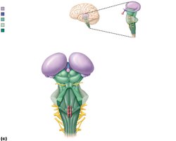
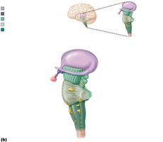
The brainstem consists of the midbrain, pons, and medulla oblongata. It is responsible for basic life functions and serves as a conduit for information between the brain and spinal cord. 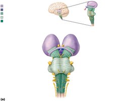
Midbrain
- Cerebral peduncles: Conduction tracts. - Corpora quadrigemina: Superior colliculi (visual reflex center), inferior colliculi (auditory relay center). - Substantia nigra: Involved in motor control. - Origin of cranial nerves: Oculomotor (III), trochlear (IV). 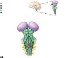
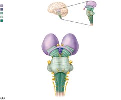
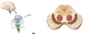
Pons
- Conduction tracts: Longitudinal and transversal fibers. - Cerebellar peduncles: Connect pons to cerebellum. - Origin of cranial nerves: Trigeminal (V), abducens (VI), facial (VII). 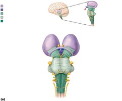
Medulla Oblongata
- Pyramids: Conduction tracts for motor signals. - Nuclei: Cuneatus, gracilis, vestibular, cochlear. - Origin of cranial nerves: Vestibulocochlear (VIII), glossopharyngeal (IX), vagus (X), hypoglossal (XII). 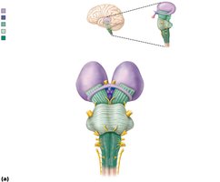
Autonomic (Visceral) Reflex Centers
- Cardiovascular center: Regulates heart rate and blood vessel diameter. - Respiratory center: Controls respiratory rhythm. - Other centers: Vomiting, hiccupping, swallowing, coughing, sneezing.
The Cerebellum
Structure and Function
The cerebellum accounts for 11% of brain mass and is essential for coordination and balance. - Processes input from cortex, brainstem, and sensory receptors. - Peduncles: Superior, middle, and inferior connect cerebellum to other brain regions. - Arbor vitae: Tree-like arrangement of white matter. 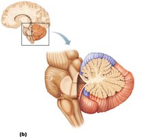
Cerebellar Processing
- Coordinates and fine-tunes motor activity: Ensures smooth, balanced skeletal muscle movement. - Receives impulses from cerebral cortex and proprioceptors. - Calculates best way to coordinate contraction and sends "blueprint" to cerebral motor cortex and brainstem nuclei.
Summary Table: Major Brain Regions and Functions
Region | Main Function |
|---|---|
Cerebrum | Conscious thought, voluntary movement, sensory processing |
Diencephalon | Relay sensory information, homeostasis, endocrine regulation |
Brainstem | Basic life functions, cranial nerve origin, conduction pathways |
Cerebellum | Coordination, balance, fine motor control |
