 Indietro
Chapter 12: The Heart – Structure, Function, and Dynamics
Guida di studio - Note intelligenti
 Appunti personalizzati basati sui tuoi materiali, ampliati con definizioni chiave, esempi e contesto.
Ch 12-1 The Heart
Anatomical Location and Orientation of the Heart
The heart is a muscular organ located in the thoracic cavity, slightly left of the midline, within the mediastinum. It is oriented so that its apex points downward and to the left, while its base is directed upward and to the right.
Location: Between the lungs, behind the sternum, and above the diaphragm.
Orientation: The apex is inferior and leftward; the base is superior and rightward.
Pericardium: The heart is enclosed in a double-layered sac called the pericardium.
Functions of the Heart
The heart functions as a pump to circulate blood throughout the body, delivering oxygen and nutrients while removing waste products.
Pulmonary Circuit: Pumps blood to the lungs for oxygenation.
Systemic Circuit: Pumps oxygenated blood to the rest of the body.
Maintains Blood Pressure: Ensures adequate perfusion of tissues.
Layers of the Heart Wall
The heart wall consists of three main layers, each with distinct structure and function:
Epicardium: The outermost layer, composed of connective tissue and fat, which protects the heart.
Myocardium: The thick, muscular middle layer responsible for contraction and pumping action.
Endocardium: The innermost layer, lining the chambers and valves, composed of endothelial cells.
Unique Features of Cardiac Muscle Cells
Cardiac muscle cells possess specialized features that enable their function:
Intercalated Discs: Specialized junctions that allow rapid transmission of electrical impulses and strong mechanical connection.
Autorhythmicity: Ability to generate their own action potentials without external stimulation.
Striations: Similar to skeletal muscle, indicating organized contractile proteins.
Branched Fibers: Allow for coordinated contraction.
Internal Structures of the Heart
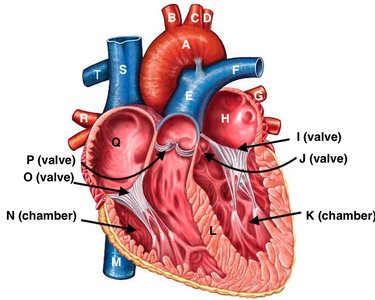
The heart contains four chambers, major blood vessels, and valves that regulate blood flow. The diagram below illustrates these internal structures:
Chambers: Right atrium, right ventricle, left atrium, left ventricle
Valves: Tricuspid, pulmonary, mitral (bicuspid), aortic
Major Vessels: Superior/inferior vena cava, pulmonary arteries/veins, aorta
Blood Flow Through the Heart
Blood flows through the heart in a specific sequence, passing through chambers, valves, and vessels:
Deoxygenated blood enters the right atrium via the superior and inferior vena cava.
Blood passes through the tricuspid valve into the right ventricle.
Right ventricle pumps blood through the pulmonary valve into the pulmonary arteries, leading to the lungs.
Oxygenated blood returns via pulmonary veins to the left atrium.
Blood passes through the mitral (bicuspid) valve into the left ventricle.
Left ventricle pumps blood through the aortic valve into the aorta, supplying the systemic circuit.
Oxygen Content in Heart Chambers and Vessels
T and F: Contain oxygen-poor blood, coming from the body and heading to the lungs.
G and R: Contain oxygen-rich blood, coming from the lungs and heading to the body.
Wall Thickness of Heart Chambers
The muscle around chamber K (left ventricle) is thicker than around chamber N (right ventricle) because the left ventricle must generate higher pressure to pump blood throughout the systemic circuit, whereas the right ventricle only pumps blood to the lungs.
Ch 12-2 Contractile Cells and Conducting System
Autorhythmicity of Heart Muscle
Autorhythmicity refers to the heart's ability to generate its own rhythmic contractions without external nervous stimulation. This is due to specialized pacemaker cells.
Pacemaking Cells and Their Location
Sinoatrial (SA) Node: Located in the right atrium; primary pacemaker of the heart.
Atrioventricular (AV) Node: Located at the junction of atria and ventricles; secondary pacemaker.
Spread of Electrical Activity in the Conducting System
The electrical impulse for heart contraction follows a specific pathway:
Impulse originates at the SA node.
Spreads through atria to the AV node.
Travels down the AV bundle (Bundle of His).
Branches into right and left bundle branches.
Reaches Purkinje fibers, causing ventricular contraction.
Electrocardiogram (ECG) Interpretation
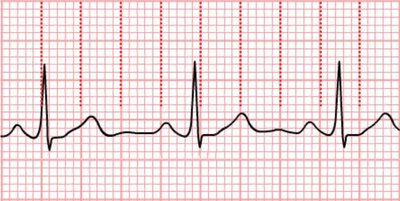
An ECG measures the electrical activity of the heart, providing information about heart rhythm, rate, and conduction abnormalities. It is useful for diagnosing arrhythmias, myocardial infarction, and other cardiac conditions.
Heart Rate Calculation from ECG
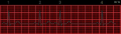
Heart rate can be calculated by counting the number of QRS complexes in a given time interval. For example, if there are 4 QRS complexes in 4 seconds, the heart rate is:
Heart rate = (Number of QRS complexes / Time in seconds) × 60
Example:
Ch 12-4 Heart Dynamics
Definitions
Cardiac Output (CO): The volume of blood pumped by the heart per minute.
Heart Rate (HR): The number of heartbeats per minute.
Stroke Volume (SV): The volume of blood ejected by each ventricle per beat.
Autonomic Effects on Heart Rate
The autonomic nervous system regulates heart rate:
Sympathetic Stimulation: Increases heart rate and force of contraction.
Parasympathetic Stimulation: Decreases heart rate.
Cardiac Output Calculations and Examples
At Rest: HR = 65 bpm, SV = 70 mL/beat.
During Race: HR = 150 bpm, SV = 110 mL/beat.
Trauma Patient: HR = 120 bpm, SV = 30 mL/beat.
Changes in Cardiac Output
Cardiac output increases during exercise due to increased heart rate and stroke volume. In trauma, heart rate may increase to compensate for decreased stroke volume caused by blood loss.
Heart Rate Increase: Due to sympathetic activation.
Stroke Volume Decrease: Due to reduced blood volume.
Summary Table: Cardiac Output Scenarios
Scenario | Heart Rate (bpm) | Stroke Volume (mL/beat) | Cardiac Output (mL/min) |
|---|---|---|---|
Rest | 65 | 70 | 4550 |
Exercise | 150 | 110 | 16500 |
Trauma | 120 | 30 | 3600 |
Additional info: Cardiac output is a critical indicator of cardiovascular health and is influenced by both intrinsic and extrinsic factors, including physical activity, autonomic regulation, and pathological conditions.
