 Indietro
Chapter 13: The Spinal Cord, Spinal Nerves, and Spinal Reflexes – Study Guide
Guida di studio - Note intelligenti
 Appunti personalizzati basati sui tuoi materiali, ampliati con definizioni chiave, esempi e contesto.
Chapter 13: The Spinal Cord, Spinal Nerves, and Spinal Reflexes
Basic Structural and Organizational Characteristics of the Nervous System
The nervous system is a complex network responsible for transmitting signals between different parts of the body. It is divided into the central nervous system (CNS) and peripheral nervous system (PNS). The spinal cord is a major CNS structure, serving as a conduit for sensory and motor information.
Central Nervous System (CNS): Includes the brain and spinal cord.
Peripheral Nervous System (PNS): Composed of nerves and ganglia outside the CNS.
Reflexes: Rapid, involuntary responses to stimuli, often processed at the spinal cord level.
Bilateral Symmetry: The spinal cord is divided into left and right halves by grooves.
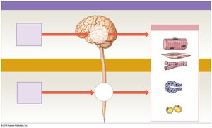
Structure and Functions of the Spinal Cord
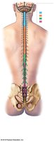
The spinal cord is housed within protective membranes (meninges) and the vertebral column. It carries sensory and motor information between the brain and most other parts of the body. The spinal cord is organized into four regions: cervical, thoracic, lumbar, and sacral, and consists of 31 segments, each giving rise to a pair of spinal nerves.
Cervical Enlargement: Supplies the shoulder and upper limb.
Lumbosacral Enlargement: Supplies the pelvis and lower limb.
Conus Medullaris: Tapered, conical end of the cord below the lumbar enlargement.
Cauda Equina: Nerve roots extending below the conus medullaris, resembling a horse’s tail.
Filum Terminale: Thin thread of fibrous tissue attaching the conus medullaris to the coccygeal ligament.
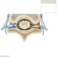
The Three Meningeal Layers
The spinal cord is protected by three meningeal layers:
Dura Mater: Outermost, tough layer.
Arachnoid Mater: Middle, web-like layer.
Pia Mater: Innermost, delicate layer adhering to the spinal cord.
Subarachnoid Space: Contains cerebrospinal fluid (CSF).
Denticulate Ligaments: Extensions of pia mater anchoring the cord to dura mater.
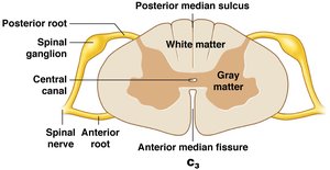
White Matter and Gray Matter in the Spinal Cord
The spinal cord contains both white and gray matter, each with distinct functions:
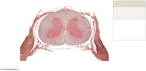
Gray Matter: Contains cell bodies of neurons, neuroglia, and unmyelinated axons. Organized into horns (posterior, anterior, lateral).
White Matter: Contains myelinated and unmyelinated axons. Organized into columns (posterior, anterior, lateral).
Commissures: Bands of gray or white matter where axons cross from one side to the other.
Functional Groups: Sensory nuclei (posterior horns), motor nuclei (anterior horns), visceral motor nuclei (lateral horns).
Spinal Nerves and Nerve Plexuses
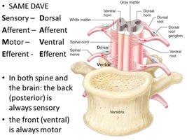
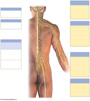
Spinal nerves are mixed nerves formed by the union of anterior (motor) and posterior (sensory) roots. Each nerve divides into rami, which supply different regions of the body. Nerve plexuses are networks of intersecting nerves that innervate specific regions.
Anterior (Ventral) Root: Contains motor (efferent) fibers.
Posterior (Dorsal) Root: Contains sensory (afferent) fibers.
Spinal Ganglia: Contain cell bodies of sensory neurons.
Rami Communicantes: Branches that innervate glands and smooth muscle.
Nerve Plexuses: Cervical, brachial, lumbar, and sacral plexuses.
Peripheral Nerve Structure
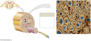
Peripheral nerves are composed of bundles of axons surrounded by connective tissue layers:
Epineurium: Outermost layer covering the nerve.
Perineurium: Surrounds each fascicle.
Endoneurium: Surrounds individual axons.
Schwann Cells: Provide myelination in the PNS.
Neuronal Pools and Patterns of Interaction
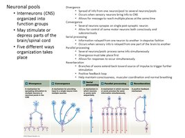
Neuronal pools are groups of interneurons organized to process information. They can stimulate or depress parts of the CNS and are organized in five main ways:
Divergence: Spreads information from one neuron to several.
Convergence: Several neurons synapse on a single neuron.
Serial Processing: Information relayed in a stepwise manner.
Parallel Processing: Several pathways process the same information simultaneously.
Reverberation: Collateral branches extend back toward the source, creating a feedback loop.
Spinal Reflexes and Reflex Arcs
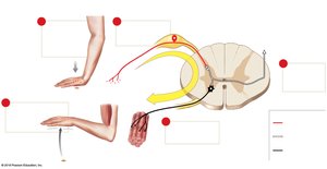
Spinal reflexes are rapid, involuntary responses processed in the spinal cord. The reflex arc is the basic functional unit of the nervous system, consisting of five steps:
Arrival of stimulus and activation of receptor
Activation of a sensory neuron
Information processing in the CNS
Activation of a motor neuron
Response by a peripheral effector
Classification of Reflexes
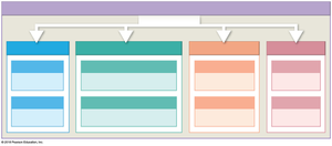
Reflexes can be classified by development, response, complexity, and processing site:
Development: Innate (genetically determined) or acquired (learned).
Response: Somatic (skeletal muscle) or visceral (autonomic, smooth/cardiac muscle, glands).
Complexity: Monosynaptic (one synapse) or polysynaptic (multiple synapses).
Processing Site: Spinal or cranial.
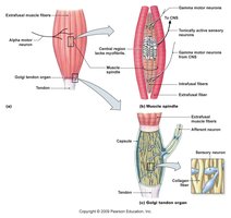
Monosynaptic Reflexes: Stretch Reflex
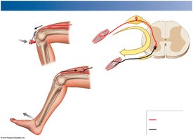
The stretch reflex is a monosynaptic reflex that regulates skeletal muscle length. It is rapid and occurs via muscle spindle receptors.
Example: Patellar (knee-jerk) reflex.
Muscle Spindle: Proprioceptors that detect changes in muscle length.
Alpha-Gamma Coactivation: Protects muscle fibers from overstretching.
Polysynaptic Reflexes
Polysynaptic reflexes involve interneurons and can control multiple muscle groups. They produce excitatory or inhibitory postsynaptic potentials (EPSPs or IPSPs).
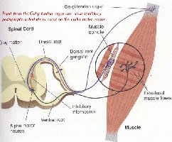
Tendon Reflex: Prevents excessive tension in muscles and tendons. Sensory receptors are Golgi tendon organs.
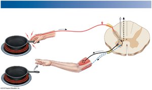
Withdrawal Reflex: Moves a body part away from a painful stimulus.
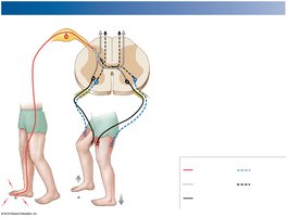
Crossed-Extensor Reflex: Coordinates opposite limb responses during withdrawal.
Characteristics of Polysynaptic Reflexes
Polysynaptic reflexes have several general characteristics:
Involve pools of interneurons
May cause excitation or inhibition
Involve more than one spinal segment
Can activate muscles in multiple areas
Involve reciprocal inhibition
Have reverberating circuits
Several reflexes may cooperate for coordinated responses
Higher Brain Centers and Reflex Modulation
Higher brain centers can control and modify reflex responses, allowing for complex behaviors and adaptation.
Plantar Reflex and Babinski Reflex
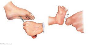
The plantar reflex is a curling of the toes seen in healthy adults. The Babinski reflex, which involves toe extension, is normal in infants but pathological in adults, indicating a lack of descending inhibition.
Summary Table: Classification of Reflexes
Classification | Type | Description |
|---|---|---|
Development | Innate | Genetically determined |
Development | Acquired | Learned |
Response | Somatic | Controls skeletal muscle contractions |
Response | Visceral | Controls actions of smooth/cardiac muscles, glands |
Complexity | Monosynaptic | One synapse |
Complexity | Polysynaptic | Multiple synapses |
Processing Site | Spinal | Processed in spinal cord |
Processing Site | Cranial | Processed in brain |
Additional info: Academic context was added to clarify the structure and function of the spinal cord, spinal nerves, and reflexes, as well as to provide definitions and examples for key terms.
