 Indietro
Chapter 14: The Brain and Cranial Nerves – Structured Study Notes
Guida di studio - Note intelligenti
 Appunti personalizzati basati sui tuoi materiali, ampliati con definizioni chiave, esempi e contesto.
The Brain and Cranial Nerves
Major Brain Regions, Vesicles, and Ventricles
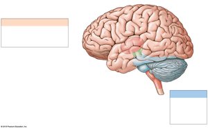
The brain is divided into several major regions, each with distinct anatomical features and functions. Understanding these regions is essential for grasping the organization and operation of the central nervous system.
Cerebrum: Responsible for conscious thought, intellectual functions, memory storage, and processing. It regulates both conscious and subconscious skeletal muscle contractions.
Cerebellum: Coordinates complex somatic motor patterns and adjusts output of other motor centers in the brain and spinal cord.
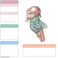
Diencephalon: Includes the thalamus (sensory relay and processing) and hypothalamus (emotions, autonomic functions, hormone production).
Brainstem: Composed of the midbrain (visual/auditory processing, reflexive motor responses, consciousness), pons (sensory relay, motor centers), and medulla oblongata (autonomic regulation, sensory relay).
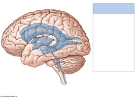
Ventricular System: Four interconnected cavities (lateral, third, fourth ventricles, and central canal) produce and circulate cerebrospinal fluid (CSF).
Brain Protection and Support
The brain is protected and supported by several physical and biochemical mechanisms.
Bones of the cranium: Provide rigid protection.
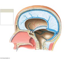
Cranial meninges: Three layers (dura mater, arachnoid mater, pia mater) surround the brain; inflammation causes meningitis.
Cerebrospinal fluid (CSF): Cushions neural structures, supports the brain, and transports nutrients, messengers, and wastes.
Blood-brain barrier: Formed by tight junctions between capillary endothelial cells; restricts passage of most substances except lipid-soluble compounds.
Anatomical Differences: Medulla Oblongata vs. Spinal Cord
The medulla oblongata is the most inferior part of the brainstem and differs from the spinal cord in both structure and function.
Medulla oblongata: Contains autonomic centers for cardiovascular and respiratory regulation, relays sensory information to the thalamus and other brainstem regions.
Spinal cord: Primarily conducts signals between the brain and peripheral nervous system.
Main Components and Functions of the Pons
The pons is a prominent structure in the brainstem, acting as a relay center and housing important motor and sensory nuclei.
Relay of sensory information: To cerebellum and thalamus.
Subconscious motor centers: For somatic and visceral functions.
Main Components and Functions of the Midbrain
The midbrain is involved in processing visual and auditory data, generating reflexive motor responses, and maintaining consciousness.
Superior colliculi: Visual reflexes.
Inferior colliculi: Auditory reflexes.
Reticular formation: Maintains alertness.
The Cerebellum
Anatomy and Functions
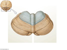
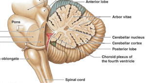
The cerebellum is essential for motor coordination and balance. It consists of two hemispheres, anterior and posterior lobes, and the vermis.
Folia: Folds of the cerebellar cortex.
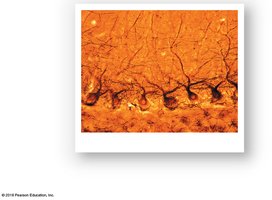
Purkinje cells: Large neurons in the cerebellar cortex, crucial for motor coordination.
Arbor vitae: Tree-like arrangement of white matter.
Disorders: Ataxia (loss of coordination) can result from trauma, stroke, or intoxication.
The Diencephalon
Thalamus
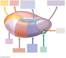
The thalamus acts as a relay and processing center for sensory information and communication between basal nuclei and the cerebral cortex.
Medial and lateral nuclei: Relay sensory and motor signals.
Geniculate bodies: Process auditory and visual input.
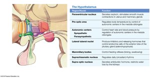
Hypothalamus
The hypothalamus regulates emotions, autonomic functions, and hormone production. It connects to the pituitary gland and controls many homeostatic processes.
Infundibulum: Connects hypothalamus to pituitary gland.
Optic chiasm: Site of visual pathway crossing.
The Limbic System
Components and Functions
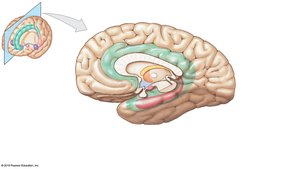
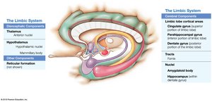
The limbic system is a group of structures involved in emotion, memory, and motivation. It links conscious and unconscious functions and facilitates memory storage and retrieval.
Cingulate gyrus: Emotional processing.
Hippocampus: Memory formation.
Mammillary bodies: Memory and olfactory processing.
Fornix: White matter tract connecting hippocampus and hypothalamus.
The Cerebrum
Anatomical Subdivisions and Functions
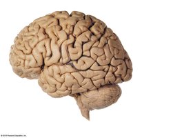
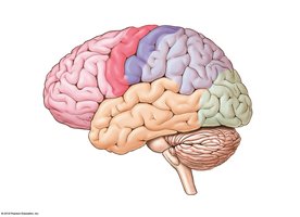
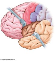
The cerebrum is divided into lobes, each with specialized functions. It is responsible for higher-order functions, including reasoning, language, and sensory perception.
Frontal lobe: Motor control, decision making, speech production.
Parietal lobe: Sensory perception, spatial processing.
Occipital lobe: Visual processing.
Temporal lobe: Auditory and olfactory processing, memory.
Insula: Emotional and limbic-related functions.
Brain Waves and Electroencephalogram (EEG)
Brain electrical activity is measured by EEG, which records different types of brain waves associated with various states of consciousness.
Alpha waves: Normal resting adults.
Beta waves: Intense concentration.
Theta waves: Children and frustrated adults.
Delta waves: Deep sleep and certain pathological conditions.
Hemispheric Lateralization
Each cerebral hemisphere specializes in certain functions. The left hemisphere is dominant for language, math, and logic, while the right hemisphere is involved in spatial abilities, face recognition, and emotional processing.
Contralateral control: Each hemisphere controls the opposite side of the body.
Ipsilateral control: Cerebellum controls the same side.
Motor, Sensory, and Association Areas
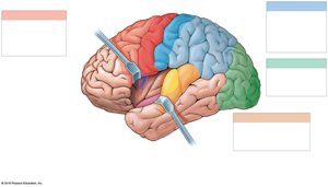
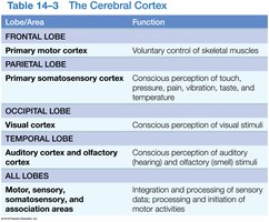
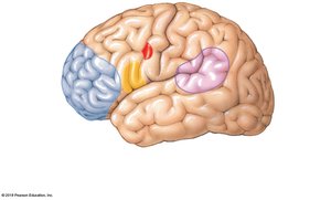
The cerebral cortex contains distinct areas for motor, sensory, and association functions.
Primary motor cortex: Voluntary control of skeletal muscles.
Primary somatosensory cortex: Perception of touch, pressure, pain, vibration, temperature.
Visual cortex: Perception of visual stimuli.
Auditory and olfactory cortex: Perception of auditory and olfactory stimuli.
Association areas: Integration and processing of sensory and motor information.
Cranial Nerves
Overview and Functions
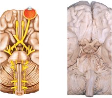
Cranial nerves are twelve pairs of nerves that emerge directly from the brain, each with specific sensory, motor, or mixed functions.
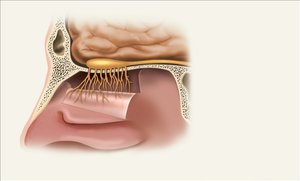
CN I: Olfactory Nerve – Sense of smell.
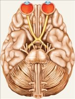
CN II: Optic Nerve – Vision.
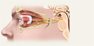
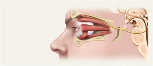
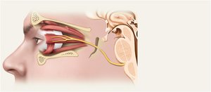
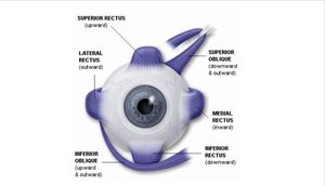
CN III: Oculomotor Nerve – Eye movement, pupil constriction.
CN IV: Trochlear Nerve – Eye movement (superior oblique muscle).
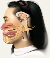
CN V: Trigeminal Nerve – Facial sensation, mastication.
CN VI: Abducens Nerve – Eye movement (lateral rectus muscle).
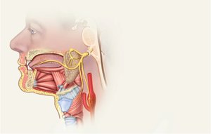
CN VII: Facial Nerve – Facial expression, taste (anterior 2/3 tongue).
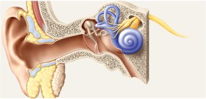
CN VIII: Vestibulocochlear Nerve – Hearing and balance.
CN IX: Glossopharyngeal Nerve – Taste (posterior 1/3 tongue), swallowing, BP regulation.
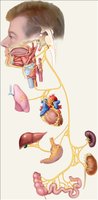
CN X: Vagus Nerve – Control of cardiac, pulmonary, digestive, urinary functions.
CN XI: Accessory Nerve – Head, neck, shoulder movement.
CN XII: Hypoglossal Nerve – Tongue movement.
Cranial Reflexes
Cranial reflexes involve sensory and/or motor fibers of cranial nerves and are clinically useful for assessing nerve and brain function.
Monosynaptic reflex: Direct connection between sensory and motor neurons.
Polysynaptic reflex: Involves interneurons.
Summary Table: Cranial Nerves
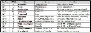
The following table summarizes the cranial nerves, their location, and function:
Number | S/M/B | Name | Location | Function |
|---|---|---|---|---|
CN I | S | Olfactory | Telencephalon | Smell - Special Sense |
CN II | S | Optic | Diencephalon | Vision - Special Sense |
CN III | M | Oculomotor | Midbrain | Eye movement (up/down/pupil) |
CN IV | M | Trochlear | Midbrain | Eye movement (superior oblique) |
CN V | B | Trigeminal | Pons | Facial Sensory and muscles of mastication |
CN VI | M | Abducent | Pons/Medulla Junction | Eye movement (lateral rectus) |
CN VII | B | Facial | Pons/Medulla Junction | Motor and intermediate nerve |
CN VIII | S | Vestibulocochlear | Pons/Medulla Junction | Hearing - Special Sense |
CN IX | B | Glossopharyngeal | Medulla | M, S, and Special Sense |
CN X | B | Vagus | Medulla | M, S, and Special Sense |
CN XI | M | Spinal Accessory | Superior Spinal Cord | Neck muscles movement |
CN XII | M | Hypoglossal | Brainstem/Medulla | Tongue muscles movement |
Example: Eye Muscles and Cranial Nerves
The extrinsic eye muscles are controlled by three cranial nerves: CN III (Oculomotor), CN IV (Trochlear), and CN VI (Abducens). The mnemonic [SO4LR6]3 helps remember: Superior Oblique by CN IV, Lateral Rectus by CN VI, all others by CN III.
Additional info:
Some details and explanations were expanded for academic completeness and clarity.
Images were included only when directly relevant to the adjacent content.
