 Indietro
Chapter 14: The Brain and Cranial Nerves – Anatomy & Physiology Study Guide
Guida di studio - Note intelligenti
 Appunti personalizzati basati sui tuoi materiali, ampliati con definizioni chiave, esempi e contesto.
The Brain and Cranial Nerves
Major Brain Regions, Vesicles, and Ventricles
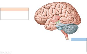
The brain is divided into several major regions, each with distinct anatomical structures and functions. Understanding these regions is essential for grasping the complexity of neural processing and integration.
Cerebrum: Responsible for conscious thought, intellectual functions, memory storage, and processing. It regulates both conscious and subconscious skeletal muscle contractions.
Cerebellum: Coordinates complex somatic motor patterns and adjusts output of other motor centers in the brain and spinal cord.
Diencephalon: Includes the thalamus (sensory relay and processing) and hypothalamus (emotions, autonomic functions, hormone production).
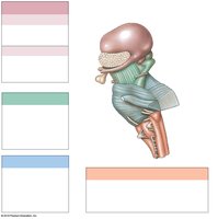
Brainstem: Composed of the midbrain (visual/auditory processing, reflexes, consciousness), pons (sensory relay, motor centers), and medulla oblongata (autonomic regulation, sensory relay).
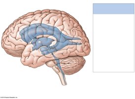
Ventricular System: Four interconnected cavities (lateral, third, fourth ventricles, and central canal) produce and circulate cerebrospinal fluid (CSF).
Protection and Support of the Brain
The brain is protected and supported by several physical and biochemical mechanisms, ensuring its integrity and function.
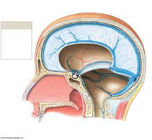
Bones of the cranium: Provide rigid protection against mechanical injury.
Cranial meninges: Three layers (dura mater, arachnoid mater, pia mater) surround the brain, offering additional protection and compartmentalization.
Cerebrospinal fluid (CSF): Cushions neural structures, supports the brain, and transports nutrients, messengers, and wastes.
Blood-brain barrier: Formed by tight junctions between capillary endothelial cells, it restricts passage of substances, allowing only lipid-soluble compounds and select molecules.
Anatomy and Function of the Medulla Oblongata
The medulla oblongata is the most inferior part of the brainstem, connecting the brain to the spinal cord. It contains vital autonomic centers and relay stations for sensory and motor pathways.
Main components: Pyramids, olives, and various nuclei.
Functions: Regulation of cardiovascular and respiratory systems, relay of sensory information to the thalamus, and integration of reflexes.
Main Components and Functions of the Pons
The pons is a prominent bulge on the brainstem, acting as a bridge between various parts of the nervous system.
Main components: Pontine nuclei, tracts, and cranial nerve nuclei.
Functions: Relays sensory information to the cerebellum and thalamus, houses motor centers for somatic and visceral functions.
Main Components and Functions of the Midbrain
The midbrain is the superior portion of the brainstem, involved in processing sensory information and generating motor responses.
Main components: Superior and inferior colliculi, substantia nigra, red nucleus.
Functions: Visual and auditory processing, reflexive motor responses, maintenance of consciousness.
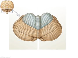
Main Components and Functions of the Cerebellum
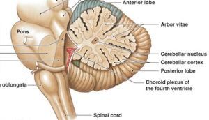
The cerebellum is located posterior to the brainstem and is essential for motor coordination and balance.
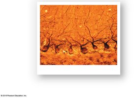
Main components: Anterior and posterior lobes, vermis, folia, cerebellar cortex, arbor vitae, Purkinje cells.
Functions: Coordination of voluntary movements, maintenance of posture and balance, fine-tuning of motor activity.
Disorders: Ataxia – disturbance in muscular coordination due to trauma, stroke, or intoxication.
Main Components and Functions of the Diencephalon
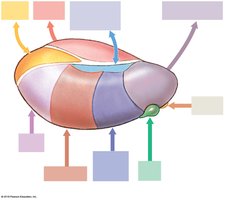
The diencephalon is a central brain region containing the thalamus and hypothalamus, which are critical for sensory processing and homeostatic regulation.
Thalamus: Filters and relays sensory information from the spinal cord and cranial nerves to the cerebral cortex; relays information between basal nuclei and cortex.
Hypothalamus: Controls emotions, autonomic functions, hormone production, and links nervous and endocrine systems.
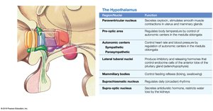
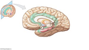
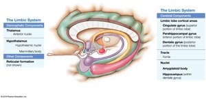
Main Components and Functions of the Limbic System
The limbic system is a group of interconnected structures involved in emotion, motivation, and memory.
Components: Cingulate gyrus, hippocampus, parahippocampal gyrus, hypothalamus, mammillary bodies, fornix, amygdaloid body.
Functions: Establishes emotional states, links conscious and unconscious functions, facilitates memory storage and retrieval.
Anatomical Subdivisions and Functions of the Cerebrum
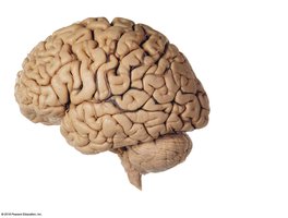
The cerebrum is the largest part of the brain, divided into hemispheres and lobes, each with specialized functions.
Lobes: Frontal, parietal, occipital, temporal, and insula.
Gyri: Ridges of nervous tissue; Sulci: Shallow grooves between gyri.
Hemispheric lateralization: Left hemisphere – language, math, decision making; Right hemisphere – sensory analysis, recognition of faces and voice inflections.
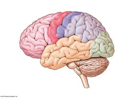
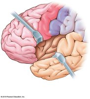
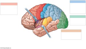
Motor, Sensory, and Association Areas of the Cerebral Cortex
The cerebral cortex contains distinct areas responsible for motor control, sensory perception, and higher-order association functions.
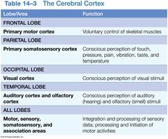
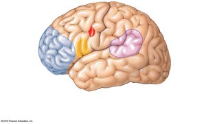
Primary motor cortex: Voluntary control of skeletal muscles (frontal lobe).
Primary somatosensory cortex: Perception of touch, pressure, pain, vibration, temperature (parietal lobe).
Visual cortex: Perception of visual stimuli (occipital lobe).
Auditory and olfactory cortex: Perception of auditory and olfactory stimuli (temporal lobe).
Association areas: Integration and processing of sensory and motor information.
Brain Waves and Electroencephalogram (EEG)
Brain electrical activity is assessed using an EEG, which records characteristic patterns known as brain waves.
Alpha waves: Normal resting adults.
Beta waves: Intense concentration.
Theta waves: Children and frustrated adults.
Delta waves: Deep sleep and certain pathological conditions.
Seizure: Temporary cerebral disorder due to abnormal electrical activity.
Cranial Nerves: Overview and Functions
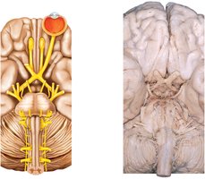
Cranial nerves are twelve pairs of nerves that emerge directly from the brain, each with specific sensory, motor, or mixed functions.
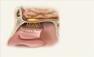
CN I: Olfactory Nerve – Sense of smell; damage causes impaired smell.
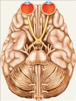
CN II: Optic Nerve – Vision; damage causes blindness.
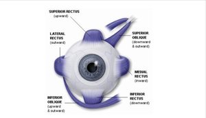
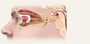
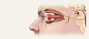
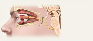
CN III: Oculomotor Nerve – Eye movement, iris, lens, eyelid; damage causes drooping eyelid, double vision.
CN IV: Trochlear Nerve – Eye movement (superior oblique); damage causes double vision.
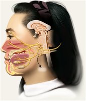
CN V: Trigeminal Nerve – Sensory/motor of face; three divisions (ophthalmic, maxillary, mandibular).
CN VI: Abducens Nerve – Eye movement (lateral rectus); damage causes inability to rotate eye laterally.
CN VII: Facial Nerve – Motor (facial expression), sensory (taste); damage causes sagging muscles, disturbed taste.
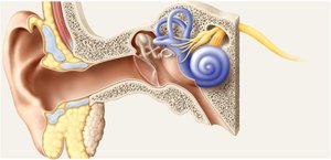
CN VIII: Vestibulocochlear Nerve – Hearing and equilibrium; damage causes deafness, dizziness.
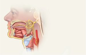
CN IX: Glossopharyngeal Nerve – Swallowing, taste, BP regulation; damage causes impaired swallowing.
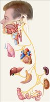
CN X: Vagus Nerve – Extensive distribution; controls cardiac, pulmonary, digestive, urinary functions.
CN XI: Accessory Nerve – Swallowing, head, neck, shoulder movement.
CN XII: Hypoglossal Nerve – Tongue movements; damage causes deviation and atrophy.
Representative Cranial Reflexes
Cranial reflexes involve sensory and/or motor fibers of cranial nerves and are clinically useful for assessing nerve and brain function.
Monosynaptic and polysynaptic reflex arcs: Simple and complex reflexes, respectively.
Examples: Pupillary reflex, corneal reflex, gag reflex.
Summary Table: Cranial Nerves
The following table summarizes the cranial nerves, their location, and primary function:
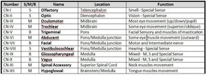
Number | S/M/B | Name | Location | Function |
|---|---|---|---|---|
CN I | S | Olfactory | Telencephalon | Smell - Special Sense |
CN II | S | Optic | Diencephalon | Vision - Special Sense |
CN III | M | Oculomotor | Midbrain | Eye movement (up/down/pupil) |
CN IV | M | Trochlear | Midbrain | Eye movement (superior oblique) |
CN V | B | Trigeminal | Pons | Facial Sensory and muscles of mastication |
CN VI | M | Abducent | Pons/Medulla Junction | Eye movement (lateral rectus) |
CN VII | B | Facial | Pons/Medulla Junction | Motor and intermediate nerve |
CN VIII | S | Vestibulocochlear | Pons/Medulla Junction | Hearing - Special Sense |
CN IX | B | Glossopharyngeal | Medulla | M, S, and Special Sense |
CN X | B | Vagus | Medulla | M, S, and Special Sense |
CN XI | M | Spinal Accessory | Superior Spinal Cord | Neck muscles movement |
CN XII | M | Hypoglossal | Brainstem/Medulla | Tongue muscles movement |
Additional info:
Some details and explanations were expanded for clarity and completeness based on standard academic context.
Images were included only when directly relevant to the adjacent content.
