 Indietro
Chapter 16: The Endocrine System – Anatomy & Physiology Study Guide
Guida di studio - Note intelligenti
 Appunti personalizzati basati sui tuoi materiali, ampliati con definizioni chiave, esempi e contesto.
Chapter 16: The Endocrine System
Overview of the Endocrine System
The endocrine system is one of the body's two major regulatory systems, alongside the nervous system. It consists of endocrine organs that synthesize and secrete hormones (chemical messengers) into the blood, which act on specific target cells containing matching receptors. Hormone binding causes functional changes in target cells, affecting various physiological processes.
Hormones: Chemical messengers released into the bloodstream.
Target Cells: Cells with specific receptors for a hormone.
Functional Changes: Hormones can stimulate/inhibit secretion, activate/inhibit enzymes, influence cell division, open/close ion channels, and change gene expression.
Comparison of the Endocrine and Nervous Systems
The nervous system uses electrical and chemical signals for rapid, short-term responses, while the endocrine system uses hormones for slower, longer-lasting regulation.
Nervous System: Fast, short-lived responses; uses neurotransmitters.
Endocrine System: Slow, long-lasting responses; uses hormones.
Paracrine and Autocrine Signals
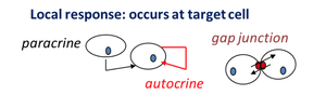
Not all cell signaling is endocrine. Paracrine signals affect nearby cells, while autocrine signals affect the same cell that secreted the signal. These are local responses and are not part of the endocrine system.
Paracrine: Local signaling to nearby cells.
Autocrine: Local signaling to self.
Gap Junctions: Direct cell-to-cell communication.
Endocrine vs. Exocrine Glands
Endocrine glands are ductless and release hormones into the interstitial fluid, which are then carried by blood. In contrast, exocrine glands use ducts to secrete substances to body surfaces or cavities.
Endocrine Glands: Ductless, release hormones into blood.
Exocrine Glands: Use ducts, secrete to surfaces/cavities.
Overview of the Endocrine Organs
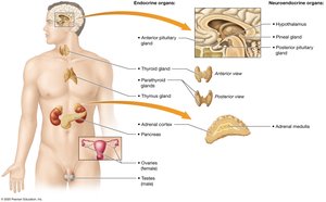
The major endocrine organs include the hypothalamus, pituitary gland, thyroid gland, parathyroid glands, adrenal glands, pancreas, pineal gland, thymus, and gonads. Each organ has specific functions and hormone secretions.
Classes of Hormones
Hormones are classified based on their chemical structure and solubility:
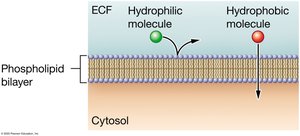
Amino Acid–Based Hormones: Mostly hydrophilic (water-soluble), except thyroid hormone (hydrophobic).
Peptide/Protein Hormones: Hydrophilic.
Steroid Hormones: Hydrophobic (lipid-soluble).
Mechanisms of Hormone Action
Hydrophilic Hormones
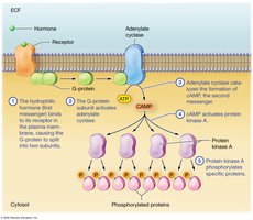
Hydrophilic hormones act via second-messenger systems, amplifying the signal inside the cell. G-proteins may activate phospholipase C, forming IP3 and DAG, or open/close ion channels.
Second Messenger: cAMP, IP3, DAG.
Amplification: One hormone can trigger many cellular responses.
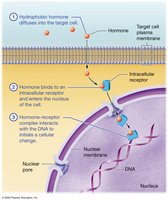
Hydrophobic Hormones
Hydrophobic hormones (steroids and thyroid hormones) bind to intracellular receptors, often affecting gene expression directly.
Intracellular Receptor: Located inside the cell, often in the nucleus.
Gene Expression: Hormone-receptor complex can initiate transcription.
Regulation of Hormone Secretion
Hormone secretion is usually regulated by negative feedback loops, maintaining homeostasis. Positive feedback is less common.
Negative Feedback: Removes the stimulus (e.g., insulin lowers high blood glucose).
Positive Feedback: Reinforces the stimulus (e.g., oxytocin during childbirth).
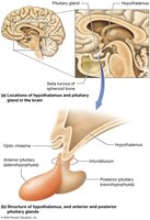
Structure of the Hypothalamus and Pituitary Gland
The hypothalamus and pituitary gland are central to endocrine regulation. The hypothalamus controls the pituitary gland, which is divided into anterior and posterior lobes.
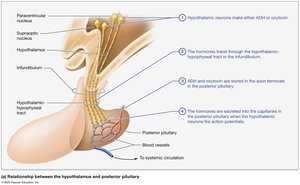
Hormones of the Hypothalamus and Posterior Pituitary
The posterior pituitary releases two main hormones:
Antidiuretic Hormone (ADH/Vasopressin): Increases water reabsorption in kidneys, raising blood volume.
Oxytocin: Causes milk ejection and uterine contraction; also acts as a bonding hormone.
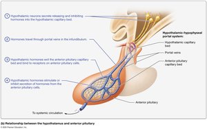
Functional Relationship of the Hypothalamus and Anterior Pituitary
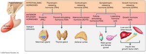
The anterior pituitary is regulated by hypothalamic releasing and inhibiting hormones. It secretes hormones that control other endocrine glands.
Hormones: Prolactin, TSH, ACTH, FSH, LH, GH.
Negative Feedback: Hormone levels regulate further secretion.
Thyroid and Parathyroid Glands
Structure of the Thyroid Gland
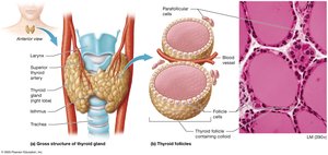
The thyroid gland is located in the anterior neck, consisting of right and left lobes joined by the isthmus. It contains follicles that produce thyroid hormones and parafollicular cells that secrete calcitonin.
Thyroid Hormones
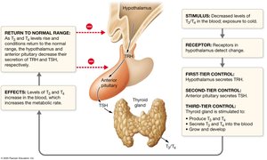
Triiodothyronine (T3): Contains three iodine atoms.
Thyroxine (T4): Contains four iodine atoms.
Functions: Regulate metabolic rate, thermoregulation, growth, and development.
Thyroid Disorders
Hyperthyroidism: Excess thyroid hormones; causes weight loss, heat intolerance, rapid heartbeat.
Graves’ Disease: Autoimmune disorder causing hyperthyroidism.
Hypothyroidism: Deficiency of thyroid hormones.
Structure of the Parathyroid Glands
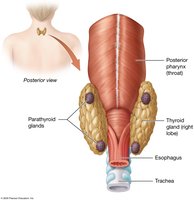
The parathyroid glands are located on the posterior surface of the thyroid gland and secrete parathyroid hormone (PTH).
Parathyroid Hormone and Calcitonin
Parathyroid Hormone (PTH): Increases blood calcium by acting on bone, kidney, and intestine.
Calcitonin: Decreases blood calcium; secreted by parafollicular cells.
Structure and Function of the Adrenal Glands
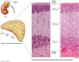
The adrenal glands are pyramid-shaped organs located on the superior end of the kidneys. They consist of the adrenal cortex and adrenal medulla.
Adrenal Cortex
Produces: Steroid hormones (cortisol, aldosterone).
Adrenal Medulla
Chromaffin Cells: Produce epinephrine and norepinephrine for the sympathetic response.
Effects: Increased heart rate, blood pressure, respiration; decreased digestive activity.
Cortisol Disorders
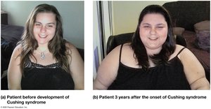
Cushing Syndrome: Excess cortisol; causes obesity, hypertension, and other symptoms.
Addison Disease: Adrenal insufficiency; hyposecretion of cortisol and aldosterone.
Structure and Function of the Pancreas
The pancreas is a club-shaped organ in the abdominal cavity, with both endocrine and exocrine functions. Endocrine cells are found in pancreatic islets, while exocrine cells (acinar cells) secrete digestive enzymes.
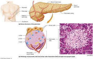
Alpha (α) Cells: Secrete glucagon; increases blood glucose.
Beta Cells: Secrete insulin; decreases blood glucose.
Delta Cells: Secrete somatostatin; inhibits GH and lowers blood glucose.
Diabetes Mellitus
Type 1: Autoimmune destruction of beta cells; requires insulin injections.
Type 2: Insulin resistance; linked to obesity and genetics; most common type.
Pineal Gland and Thymus
Pineal Gland
Location: Epithalamus of the diencephalon.
Hormone: Melatonin; regulates sleep/wake cycle.
Thymus
Location: Mediastinum.
Hormones: Thymosin and thymopoietin; assist in T lymphocyte maturation.
The Gonads: Sex Hormones
Testes (Male)
Produce: Sperm and testosterone.
FSH: Stimulates sperm development.
LH: Stimulates testosterone secretion and male traits.
Ovaries (Female)
Produce: Oocytes, estrogen, and progesterone.
FSH & LH: Control follicle growth and ovulation.
Estrogen & Progesterone: Regulate uterine cycle and female secondary traits.
Summary Table: Major Endocrine Glands and Their Hormones
Gland | Main Hormones | Main Effects |
|---|---|---|
Hypothalamus | Releasing/inhibiting hormones | Regulate pituitary |
Pituitary (anterior) | GH, TSH, ACTH, FSH, LH, PRL | Growth, metabolism, reproduction |
Pituitary (posterior) | ADH, Oxytocin | Water balance, uterine contraction |
Thyroid | T3, T4, Calcitonin | Metabolism, calcium regulation |
Parathyroid | PTH | Increase blood calcium |
Adrenal Cortex | Cortisol, Aldosterone | Stress, water/salt balance |
Adrenal Medulla | Epinephrine, Norepinephrine | Fight-or-flight response |
Pancreas | Insulin, Glucagon, Somatostatin | Blood glucose regulation |
Pineal | Melatonin | Sleep/wake cycle |
Thymus | Thymosin, Thymopoietin | T cell maturation |
Gonads | Testosterone, Estrogen, Progesterone | Reproduction, secondary traits |
Key Homeostatic Imbalances
Diabetes Mellitus: Insulin deficiency or resistance.
Addison’s Disease: Adrenal insufficiency.
Cushing’s Syndrome: Excess cortisol.
Hypothyroidism/Hyperthyroidism: Thyroid hormone imbalances.
Additional info: Academic context was added to clarify hormone mechanisms, feedback regulation, and disease states for completeness.
