 Indietro
Chapter 17: The Special Senses – Anatomy & Physiology Study Guide
Guida di studio - Note intelligenti
 Appunti personalizzati basati sui tuoi materiali, ampliati con definizioni chiave, esempi e contesto.
The Special Senses
Introduction to the Special Senses
The special senses include olfaction (smell), gustation (taste), vision, equilibrium (balance), and hearing. These senses are mediated by specialized sensory organs and provide critical information about our environment.
Olfaction: Detection of airborne chemicals.
Gustation: Detection of dissolved chemicals in food and liquids.
Vision: Detection of light and color.
Equilibrium: Maintenance of balance and spatial orientation.
Hearing: Detection of sound waves.
Olfaction (Smell)
Olfactory Organs and Pathways
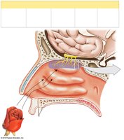
Olfactory organs are located in the nasal cavity and consist of olfactory epithelium, olfactory nerve fibers, and associated structures.
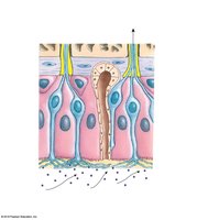
Olfactory epithelium: Contains olfactory sensory neurons, supporting cells, and basal epithelial cells.
Olfactory nerve fibers (CN I): Transmit signals through the cribriform plate to the olfactory bulb.
Olfactory bulb and tract: Relay information to the central nervous system.
Olfactory Reception and Discrimination
Olfactory receptors are modified neurons with cilia-shaped dendrites. Odorant molecules bind to receptor proteins, leading to depolarization and action potential generation.
Odorant-binding proteins: Facilitate interaction with odorants.
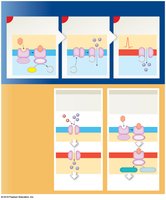
Signal transduction: Activation of adenylate cyclase, production of cAMP, opening of sodium channels, and depolarization.
Olfactory discrimination: Each receptor responds to specific odorants; as few as four molecules can activate a receptor.
Gustation (Taste)
Taste Buds and Lingual Papillae
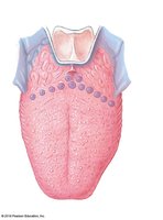
Gustatory epithelial cells are located in taste buds, which are found on lingual papillae on the tongue and parts of the pharynx and larynx.
Types of papillae: Vallate, foliate, fungiform (contain taste buds); filiform (provide friction, no taste buds).
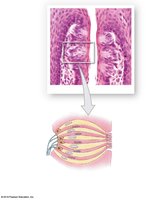
Taste buds: Contain basal epithelial cells (stem cells) and gustatory epithelial cells (receptors).
Taste hairs: Microvilli extend through taste pores to detect tastants.
Gustatory Pathways and Discrimination
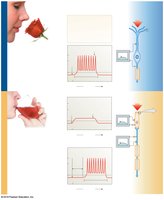
Taste information is transmitted via cranial nerves to the medulla oblongata, thalamus, and gustatory cortex of the insula.
Receptor mechanisms: Salt and sour tastes involve ion channels; sweet, bitter, and umami involve G protein-coupled receptors (gustducins).
Discrimination: Most gustatory cells respond to multiple tastants; different tastes use distinct receptor pathways.
Integration of Olfactory and Gustatory Information
Brain Regions Involved
Olfactory and gustatory cortices are located in the insula, frontal lobe, and uncus of the temporal lobe. These areas integrate sensory information for perception. 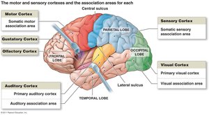
Vision
Internal and Accessory Structures of the Eye
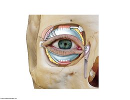
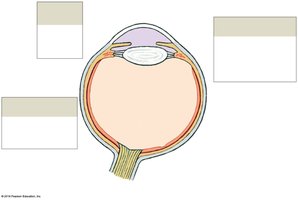
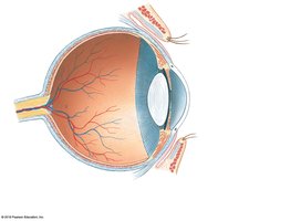
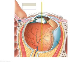
The eye consists of three main layers: fibrous (outer), vascular (middle/uvea), and inner (retina). Accessory structures include eyelids, conjunctiva, lacrimal apparatus, and extraocular muscles.
Fibrous layer: Sclera (white of eye), cornea (transparent).
Vascular layer: Iris (controls pupil diameter), ciliary body, choroid (supplies nutrients).
Inner layer: Retina (contains photoreceptors: rods and cones).
Lacrimal apparatus: Produces and drains tears.
Pupillary Muscles and Light Regulation
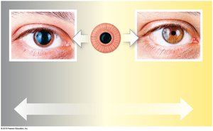
The iris contains sphincter and dilator pupillae muscles that regulate pupil diameter in response to light intensity and autonomic stimulation.
Sphincter pupillae: Constricts pupil (parasympathetic).
Dilator pupillae: Dilates pupil (sympathetic).
Retinal Organization and Photoreceptors
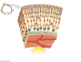
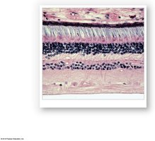
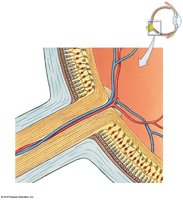
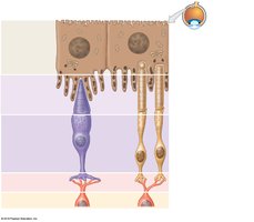
The retina contains layers of cells: photoreceptors (rods and cones), bipolar cells, ganglion cells, horizontal and amacrine cells.
Rods: Sensitive to dim light, peripheral vision.
Cones: Color vision, high visual acuity, concentrated in fovea centralis.
Horizontal and amacrine cells: Modulate signal transmission and sensitivity.
Refraction and Focusing of Light
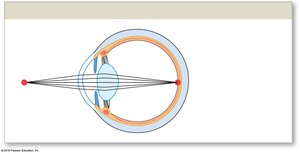
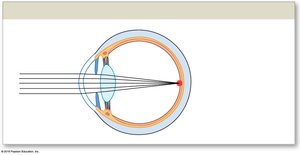
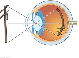
Light is refracted by the cornea and lens to focus images on the retina.
Focal point: Intersection of light rays on retina.
Focal distance: Distance from lens center to focal point.
Astigmatism: Distorted vision due to irregular lens/cornea shape.
Visual acuity: Clarity of vision (20/20 standard).
Refractive Errors
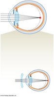
Myopia (nearsightedness): Image focused in front of retina; corrected with diverging lens.
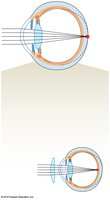
Hyperopia (farsightedness): Image focused behind retina; corrected with converging lens.
Presbyopia: Age-related loss of lens elasticity.
Photoreceptor Structure and Visual Pigments
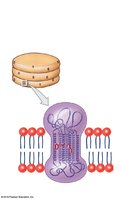
Rods: Contain rhodopsin (opsin + retinal).
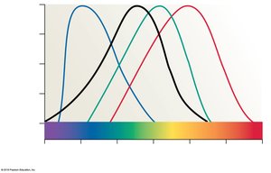
Cones: Contain photopsins (different opsins for blue, green, red).
Photobleaching: Breakdown and reassembly of visual pigments after light absorption.
Color Vision and Perception
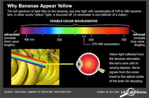
Color vision is mediated by three types of cones, each sensitive to different wavelengths.
Color blindness: Inability to distinguish certain colors due to missing cone types.
Perception: Objects appear colored based on reflected wavelengths; black absorbs all, white reflects all.
Photoreception Mechanism
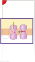
Opsin activation: Light converts 11-cis retinal to 11-trans, activating opsin.
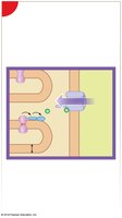
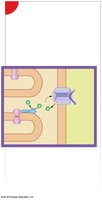
Transducin and PDE: Opsin activates transducin, which activates phosphodiesterase (PDE).
cGMP breakdown: PDE reduces cGMP, closing sodium channels and hyperpolarizing the cell.
Signal transmission: Reduced neurotransmitter release signals photon absorption.
Visual Pathways and Depth Perception
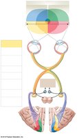
Visual information travels from photoreceptors to bipolar and ganglion cells, then via the optic nerve, optic chiasm, and optic tract to the visual cortex.
Depth perception: Comparison of images from both eyes enables 3D vision.
Crossing over: Optic chiasm allows hemispheric integration of visual fields.
The Ear: Hearing and Equilibrium
Anatomy of the Ear
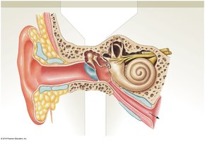
The ear is divided into external, middle, and internal regions.
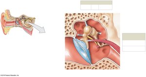
External ear: Auricle, external acoustic meatus, tympanic membrane.
Middle ear: Auditory ossicles (malleus, incus, stapes), oval and round windows, auditory tube.
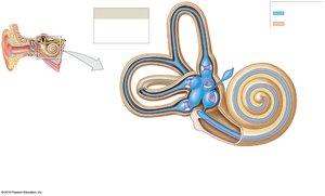
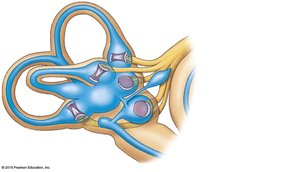
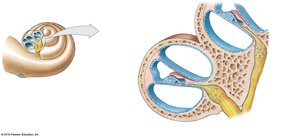
Internal ear: Bony and membranous labyrinths, cochlea, vestibule, semicircular canals.
Equilibrium: Static and Dynamic
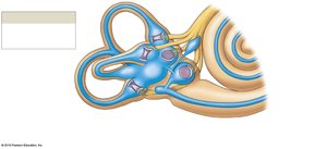
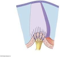
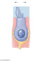
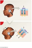
Equilibrium is maintained by the vestibular complex (vestibule and semicircular canals) and hair cells.
Semicircular ducts: Detect angular movement/rotation.
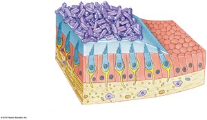
Vestibule (utricle and saccule): Detect gravity and linear acceleration.
Maculae: Clusters of hair cells sensing position and movement.
Otoliths: Calcium carbonate crystals aiding static equilibrium.
Process of Hearing
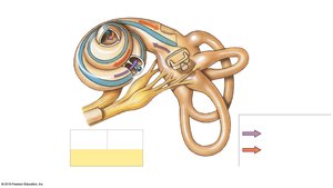
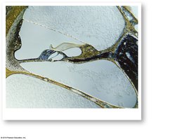
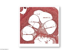
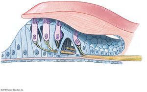
Sound waves are converted to mechanical movements, then to pressure waves in the cochlear fluid, and finally detected by hair cells in the spiral organ (organ of Corti).
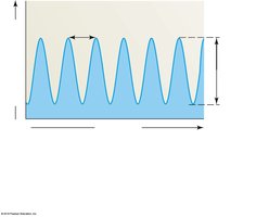
Sound properties: Frequency (pitch, Hz), amplitude (loudness, dB).
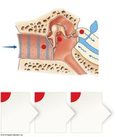
Auditory pathway: Tympanic membrane → ossicles → oval window → cochlea → spiral organ → cochlear nerve → auditory cortex.
Discrimination of Sound Frequencies
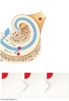
Different regions of the basilar membrane and organ of Corti are activated by different sound frequencies, enabling pitch discrimination (Place Theory).
Frequency | Basilar Membrane Region | Perceived Pitch |
|---|---|---|
High | Base (near oval window) | High pitch |
Low | Apex (far from oval window) | Low pitch |
Additional info: Outer hair cells amplify sound, inner hair cells transmit signals to the brain.
