 Indietro
Chapter 18: The Endocrine System – Anatomy & Physiology Study Notes
Guida di studio - Note intelligenti
 Appunti personalizzati basati sui tuoi materiali, ampliati con definizioni chiave, esempi e contesto.
The Endocrine System
Introduction to the Endocrine System
The endocrine system is a major regulatory system in the body, responsible for maintaining homeostasis through the release of hormones. It works in concert with the nervous system to coordinate and regulate physiological processes.
Hormones: Chemical messengers released by endocrine glands, transported via the bloodstream to target organs.
Homeostasis: The maintenance of stable internal conditions.
Intercellular Communication: Essential for coordinating cellular activities and responses.
Mechanisms of Intercellular Communication
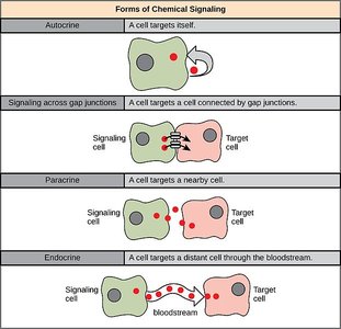
Cells communicate through several mechanisms, each with distinct characteristics and functions.
Direct Communication: Exchange of ions and molecules between adjacent cells via gap junctions; rare and highly specialized.
Paracrine Communication: Chemical signals transfer information between cells within a single tissue.
Autocrine Communication: Cells release signals that affect themselves; e.g., prostaglandins in smooth muscle.
Endocrine Communication: Endocrine cells release hormones into the bloodstream, affecting distant target cells.
Synaptic Communication: Neurons release neurotransmitters at synapses for rapid, targeted responses.
Comparison of Endocrine and Nervous Systems
Both systems rely on chemical messengers and negative feedback mechanisms to regulate activities and preserve homeostasis.
Shared Messengers: Some chemicals, such as norepinephrine and epinephrine, function in both systems.
Regulation: Negative feedback is the primary regulatory mechanism.
Function: Coordination and regulation of physiological activities.
Organs and Tissues of the Endocrine System
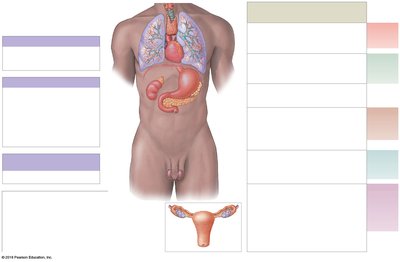
The endocrine system consists of primary glands and organs with secondary endocrine functions.
Primary Endocrine Organs: Hypothalamus, pituitary gland, thyroid gland, parathyroid glands, adrenal glands, pancreas, pineal gland.
Secondary Endocrine Functions: Heart, thymus, adipose tissue, digestive tract, kidneys, gonads (testes and ovaries).

Hormones: Structure, Transport, and Action
Hormone Transport and Inactivation
Hormones may circulate freely or be bound to carrier proteins. Their activity and duration depend on their chemical nature.
Free Hormones: Remain functional for less than an hour; inactivated by binding to target cells, breakdown in liver/kidneys, or enzymatic degradation.
Bound Hormones: Thyroid and steroid hormones are mostly bound to transport proteins, providing a reserve and longer activity.
Hormone-Receptor Interactions
The cellular response to hormones depends on the number and sensitivity of receptors.
Regulation of Receptor Number: Cells can upregulate or downregulate receptors to adjust sensitivity.
Amplification: Small amounts of hormone can trigger large cellular responses via second messengers.
Mechanisms of Hormonal Action
Hormones act through different mechanisms depending on their solubility and structure.
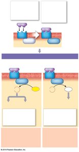
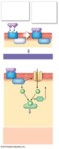
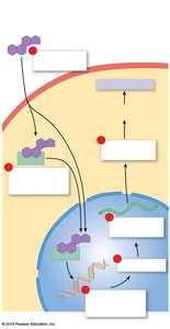
Peptide Hormones and Catecholamines: Lipid-insoluble; bind to extracellular receptors and use second messengers (e.g., cAMP, Ca2+).
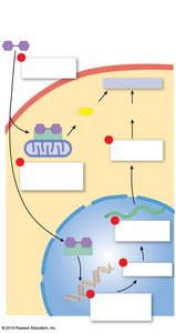
Steroid Hormones: Lipid-soluble; diffuse through membranes and bind to intracellular receptors, affecting gene expression.
Thyroid Hormones: Lipid-soluble; enter cells and bind to nuclear and mitochondrial receptors, influencing gene activation and ATP production.
The Pituitary Gland
Anatomy and Function
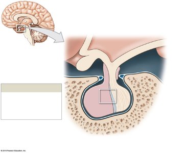
The pituitary gland, or hypophysis, is often called the 'master gland' due to its regulatory influence over other endocrine organs.
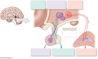
Location: Lies within the sella turcica, inferior to the hypothalamus, connected by the infundibulum.
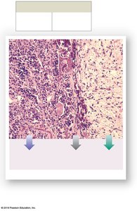
Structure: Composed of anterior (adenohypophysis) and posterior (neurohypophysis) lobes.
Hormones: Releases nine peptide hormones, most using cAMP as a second messenger.
Hypothalamic Control of Pituitary Function
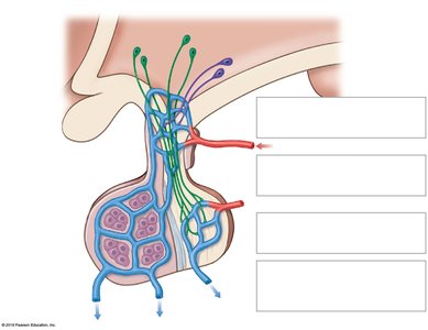
The hypothalamus regulates pituitary activity through direct neural connections and regulatory hormones.
Regulatory Hormones: Releasing hormones (RH) stimulate, inhibiting hormones (IH) suppress anterior pituitary secretion.
Portal System: Hypophyseal portal vessels ensure rapid delivery of regulatory hormones.
Pituitary Hormones and Their Targets
The pituitary gland produces hormones that regulate various target organs and tissues.
Anterior Pituitary: ACTH, TSH, GH, PRL, FSH, LH, MSH
Posterior Pituitary: ADH, OXT
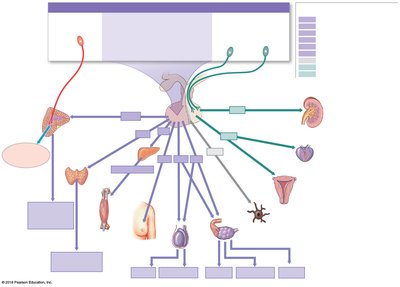
Hormone | Target Organ/Tissue | Principal Effects |
|---|---|---|
FSH | Ovaries, testes | Growth of ovarian follicles, estrogen secretion, sperm production |
LH | Ovaries, testes | Ovulation, corpus luteum maintenance, testosterone secretion |
TSH | Thyroid gland | Secretion of thyroid hormones |
ACTH | Adrenal gland | Secretion of glucocorticoids |
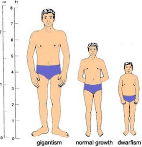
GH | Liver, bone, cartilage, muscle, fat | Widespread tissue growth |
PRL | Mammary glands, testes | Milk synthesis, LH sensitivity |
ADH | Kidneys | Water retention |
OXT | Uterus, mammary glands | Labor contraction, milk release |
The Thyroid Gland
Anatomy and Histology
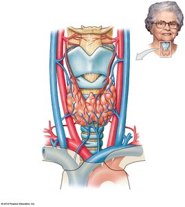
The thyroid gland is located inferior to the thyroid cartilage of the larynx and consists of two lobes connected by an isthmus.
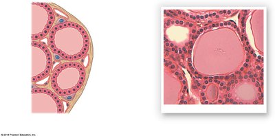
Thyroid Follicles: Hollow spheres lined by cuboidal epithelium, containing colloid.
C Cells: Parafollicular cells that produce calcitonin.
Thyroid Hormone Synthesis and Regulation
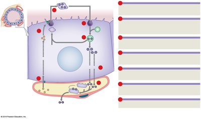
Thyroid hormones are synthesized from thyroglobulin and iodide ions, stored in follicles, and released into the bloodstream.
Thyroxine (T4): Contains four iodine atoms.
Triiodothyronine (T3): Contains three iodine atoms.
Regulation: Controlled by TSH from the pituitary gland.
Effects and Disorders of Thyroid Hormones
Thyroid hormones affect nearly every cell, regulating metabolism, growth, and development. Disorders include hyperthyroidism, hypothyroidism, and goiter.
Hyperthyroidism: Excessive TH production; symptoms include weight loss, hyperactivity, heat intolerance.
Hypothyroidism: Deficient TH production; symptoms include lethargy, weight gain, cold intolerance.
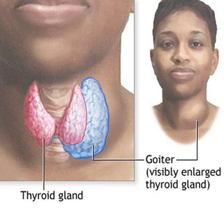
Goiter: Enlargement of the thyroid gland, often due to iodine deficiency.
Calcitonin and Calcium Regulation
Calcitonin, produced by C cells, lowers blood calcium levels by stimulating excretion and inhibiting absorption.
Stimulates: Ca2+ excretion by kidneys.
Inhibits: Ca2+ absorption in the digestive tract.
The Parathyroid Glands
Anatomy and Function
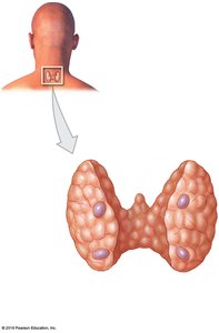
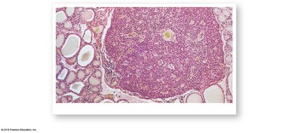
The parathyroid glands are small, paired glands located on the posterior surface of the thyroid gland. They regulate calcium homeostasis.
Parathyroid Hormone (PTH): Increases blood calcium levels by stimulating osteoclasts, enhancing kidney reabsorption, and promoting calcitriol production.
Antagonist: Calcitonin.
The Adrenal Glands
Anatomy and Hormones
The adrenal glands are located atop each kidney and consist of an outer cortex and inner medulla, each producing distinct hormones.
Adrenal Cortex: Produces corticosteroids (mineralocorticoids, glucocorticoids, and androgens).
Adrenal Medulla: Produces catecholamines (epinephrine and norepinephrine).
Adrenal Hormones and Their Effects
Region/Zone | Hormones | Primary Target | Effects |
|---|---|---|---|
Zona glomerulosa | Aldosterone | Kidneys | Increase Na+ reabsorption, water retention, K+ excretion |
Zona fasciculata | Glucocorticoids (cortisol) | Most cells | Increase glucose formation, anti-inflammatory |
Zona reticularis | Androgens | Most cells | Stimulate pubic hair development |
Medulla | Epinephrine, norepinephrine | Most cells | Increase cardiac activity, blood pressure, glucose levels |
Disorders of Adrenal Cortex Hormones
Cushing Syndrome: Excess glucocorticoids; symptoms include obesity, hypertension, and menstrual irregularities.
Addison Disease: Adrenal insufficiency; symptoms include weight loss, fatigue, hypotension.
Adrenogenital Syndrome: Excess androgen production; masculinization effects in newborns.
The Pineal Gland
Anatomy and Function
The pineal gland is located in the posterior roof of the third ventricle and produces melatonin.
Melatonin: Regulates circadian rhythms, inhibits reproductive functions, and protects against free radicals.
The Pancreas
Anatomy and Function
The pancreas is a large gland with both exocrine and endocrine functions, located between the stomach and small intestine.
Exocrine Pancreas: Produces digestive enzymes.
Endocrine Pancreas: Contains islets of Langerhans, producing insulin, glucagon, somatostatin, and pancreatic polypeptide.
Regulation of Blood Glucose
Insulin: Lowers blood glucose by promoting uptake and storage.
Glucagon: Raises blood glucose by stimulating release from storage.
Disorders Associated with the Pancreas
Diabetes Mellitus: Chronic hyperglycemia; includes Type 1 (insulin-dependent), Type 2 (insulin resistance), and gestational diabetes.
Complications: Kidney degeneration, retinal damage, heart attacks, neuropathies, tissue damage.
Hypoglycemia: Low blood glucose; symptoms include hunger, dizziness, confusion.
Secondary Endocrine Functions
Other Organs with Endocrine Roles
Kidneys: Produce erythropoietin (EPO) and renin, regulate blood pressure and red blood cell production.
Heart: Produces ANP and BNP, reduces blood volume and pressure.
Thymus: Produces thymic hormones for T-cell maturation.
Gonads: Testes produce testosterone and inhibin; ovaries produce estrogens, progesterone, and inhibin.
Adipose Tissue: Secretes leptin, regulates appetite and reproductive functions.
Hormone Interactions and General Adaptation Syndrome
Hormones Important to Growth
Growth Hormone (GH): Supports development and metabolism.
Thyroid Hormones: Essential for nervous and skeletal development.
Insulin: Facilitates glucose and amino acid uptake.
PTH and Calcitriol: Promote calcium absorption and bone deposition.
Reproductive Hormones: Stimulate growth and differentiation, influence secondary sex characteristics.
General Adaptation Syndrome (GAS)
The body's response to stress is divided into three phases:
Alarm Phase: Immediate response; epinephrine is dominant.
Resistance Phase: Prolonged stress; glucocorticoids are dominant.
Exhaustion Phase: Breakdown of homeostasis; organ failure may occur.
Aging and the Endocrine System
Secretory Activity: Decreases with age, reducing hormone levels and efficiency.
Effects: Loss of body mass, osteoporosis, changes in metabolism and reproductive function.
Integration with Other Body Systems
The endocrine system interacts with all other organ systems to regulate growth, metabolism, fluid balance, and stress responses.
Additional info: This study guide covers the major glands, hormones, mechanisms, and clinical disorders of the endocrine system, providing a comprehensive overview for Anatomy & Physiology students.
