 Indietro
Chapter 19: Blood – Structure, Function, and Clinical Relevance
Guida di studio - Note intelligenti
 Appunti personalizzati basati sui tuoi materiali, ampliati con definizioni chiave, esempi e contesto.
Blood and the Cardiovascular System
Introduction to Blood
Blood is a specialized connective tissue that plays a vital role in the cardiovascular system, which also includes the heart and blood vessels. It consists of cells suspended in a fluid matrix and is essential for transporting substances, regulating homeostasis, and defending the body.
Transport: Oxygen, carbon dioxide, nutrients, hormones, immune components, and wastes.
Regulation: pH, ion composition, and body temperature.
Protection: Restricts fluid loss, defends against toxins/pathogens.
Characteristics and Composition of Blood
Physical and Chemical Properties
Blood is a viscous fluid with a temperature of about 38°C (100.4°F).
It has a slightly alkaline pH (7.35–7.45).
Blood volume is about 7% of body weight (5–6 L in males, 4–5 L in females).
Oxygen-rich blood is bright red; oxygen-poor blood is darker red.
Components of Blood
Plasma: About 55% of blood volume; over 90% water; contains plasma proteins, nutrients, hormones, electrolytes, dissolved gases, and waste products.
Formed Elements: About 45% of blood volume; includes red blood cells (RBCs), white blood cells (WBCs), and platelets.
Plasma Proteins
Types and Functions
Albumins (60%): Maintain osmotic pressure, transport fatty acids, steroid and thyroid hormones.
Globulins (35%): Include antibodies (immunoglobulins) and transport proteins.
Fibrinogen (4%): Essential for blood clotting; converted to fibrin during coagulation.
Other Proteins (1%): Include peptide hormones and regulatory proteins (e.g., insulin, TSH).
Serum is plasma without clotting proteins (fibrinogen).
Formed Elements
Red Blood Cells (Erythrocytes)
RBCs make up 99.9% of formed elements and are specialized for oxygen transport. They are biconcave, lack nuclei and organelles, and contain hemoglobin.
Hemoglobin (Hb): Each molecule has four globular subunits, each with a heme group (iron ion). Binds oxygen (oxyhemoglobin) and carbon dioxide (carbaminohemoglobin).
Normal Hb: Males: 14–18 g/dL; Females: 12–16 g/dL.
Life Span: ~120 days; old RBCs are removed by macrophages in the liver, spleen, and bone marrow.
RBC Recycling: Hemoglobin is broken down; iron is recycled, heme is converted to biliverdin and then bilirubin, which is excreted in bile. Excess bilirubin causes jaundice.
Hematocrit
Hematocrit is the percentage of formed elements in blood (mainly RBCs). Normal values: Males 40–54%, Females 37–47%.
Low Hematocrit: May indicate anemia or blood loss.
High Hematocrit: May indicate dehydration or polycythemia.
Erythropoiesis
The process of RBC production in red bone marrow, stimulated by erythropoietin (EPO) from the kidneys during hypoxia. Requires amino acids, iron, vitamin B12, B6, and folic acid.
Pernicious anemia: Caused by vitamin B12 deficiency.
Reticulocytes: Immature RBCs released into circulation.
Blood Typing
ABO and Rh Blood Groups
Blood types are determined by the presence or absence of antigens (agglutinogens) on RBCs. Plasma contains antibodies (agglutinins) against foreign antigens.
ABO System: Types A, B, AB, O based on A and B antigens.
Rh System: Rh+ (D antigen present), Rh− (D antigen absent).
If incompatible blood is transfused, agglutination and hemolysis may occur.
Anti-A | Anti-B | Anti-Rh | Blood type |
|---|---|---|---|
Clumping | No clumping | Clumping | A+ |
No clumping | Clumping | Clumping | B+ |
Clumping | Clumping | Clumping | AB+ |
No clumping | No clumping | No clumping | O− |
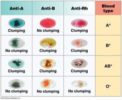
ABO Blood Group System
Type A: A antigens, anti-B antibodies; can donate to A, AB; receive from A, O.
Type B: B antigens, anti-A antibodies; can donate to B, AB; receive from B, O.
Type AB: A and B antigens, no anti-A/B antibodies; universal recipient.
Type O: No A/B antigens, both anti-A and anti-B antibodies; universal donor.
Rh Factor and Clinical Implications
Rh+: D antigen present.
Rh−: D antigen absent; anti-Rh antibodies develop after exposure.
Hemolytic Disease of the Newborn (HDN): Occurs when an Rh− mother carries an Rh+ fetus; maternal antibodies may attack fetal RBCs in subsequent pregnancies.
Transfusion Reactions: Occur with incompatible blood; can cause agglutination, hemolysis, kidney damage, and shock.
Universal Donor: O− (lacks A, B, Rh antigens).
Universal Recipient: AB+ (no antibodies against A, B, or Rh).
White Blood Cells (Leukocytes)
General Features and Functions
Have nuclei and organelles; lack hemoglobin.
Can migrate into tissues (diapedesis), respond to chemotaxis, and exhibit amoeboid movement.
Defend against pathogens, remove toxins/debris, and participate in immune responses.
Leukopoiesis
Production of WBCs in red bone marrow from hemocytoblasts. Regulated by colony-stimulating factors (CSFs) and interleukins.
Myeloid Stem Cells: Produce neutrophils, eosinophils, basophils, monocytes.
Lymphoid Stem Cells: Produce lymphocytes (T cells, B cells, NK cells).
Types of Leukocytes
Granulocytes: Visible cytoplasmic granules; include neutrophils, eosinophils, basophils.
Agranulocytes: Lack visible granules; include monocytes, lymphocytes.
Granulocytes
Neutrophils: Most abundant; first responders to bacterial infection; phagocytic.
Eosinophils: Attack parasites; increase in allergies; regulate inflammation.
Basophils: Release histamine (inflammation) and heparin (anticoagulant).
Agranulocytes
Monocytes: Largest WBCs; become macrophages in tissues; phagocytize pathogens and debris.
Lymphocytes: Main cells of specific immunity; include T cells (cell-mediated immunity), B cells (antibody production), and NK cells (destroy infected/cancer cells).
Clinical Terms
Leukopenia: Low WBC count.
Leukocytosis: High WBC count.
Leukemia: Cancer with uncontrolled WBC production.
Platelets (Thrombocytes)
Structure and Function
Small cell fragments from megakaryocytes; lack nuclei.
Essential for blood clotting (hemostasis).
Normal count: 150,000–500,000/μL.
Thrombocytopenia: Low platelet count; risk of bleeding.
Thrombocytosis: High platelet count; risk of abnormal clotting.
Platelet Production (Thrombocytopoiesis)
Occurs in red bone marrow; platelets are fragments of megakaryocytes.
Functions of Platelets
Release clotting chemicals.
Form temporary platelet plugs.
Reduce size of vessel breaks.
Hemostasis and Coagulation
Phases of Hemostasis
Vascular Phase: Vasoconstriction reduces blood loss; endothelial cells release endothelins and become sticky.
Platelet Phase: Platelets adhere to exposed collagen, aggregate, and release chemicals (ADP, serotonin, Ca2+, PDGF).
Coagulation Phase: Cascade of reactions converts fibrinogen to fibrin, forming a stable clot.
Coagulation Pathways
Extrinsic Pathway: Initiated by tissue factor (TF) from damaged tissues; activates Factor X quickly.
Intrinsic Pathway: Triggered by exposed collagen; involves platelet factors; activates Factor X through multiple steps.
Common Pathway: Factor X forms prothrombinase, which converts prothrombin to thrombin; thrombin converts fibrinogen to fibrin.
Anticoagulants and Clot Removal
Anticoagulants: Heparin, aspirin, antithrombin III inhibit clotting.
Clot Retraction: Stabilizes the clot.
Fibrinolysis: Dissolves clots via plasmin.
