 Indietro
Chapter 19: Blood Vessels – Structure, Function, and Clinical Relevance
Guida di studio - Note intelligenti
 Appunti personalizzati basati sui tuoi materiali, ampliati con definizioni chiave, esempi e contesto.
Blood Vessels: Overview and Functions
General Facts and Importance
Blood vessels form an extensive network throughout the human body, delivering oxygen and nutrients to tissues while removing metabolic waste. The total length of blood vessels in the human body is approximately 60,000 miles, with about 10 billion capillaries facilitating exchange at the tissue level. This vast network is essential for maintaining tissue metabolism and homeostasis.
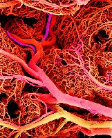
Arteries carry blood away from the heart.
Veins return blood to the heart.
Capillaries connect the smallest arteries to the smallest veins and are the primary sites of exchange.
Anatomy and Structure of Blood Vessels
Histological Structure
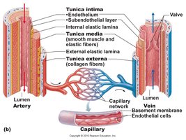
Blood vessels are composed of three main layers (tunics):
Tunica intima: Innermost layer, composed of endothelium and subendothelial connective tissue.
Tunica media: Middle layer, primarily smooth muscle and elastic fibers, responsible for vasoconstriction and vasodilation.
Tunica externa (adventitia): Outermost layer, composed of collagen fibers that protect and anchor the vessel.
Types of Blood Vessels
Arteries
Arteries are resistance vessels that withstand high blood pressure generated by ventricular contraction. They are muscular and retain a round shape when empty.
Conducting (elastic) arteries: Largest arteries (e.g., aorta), expand and recoil to maintain blood pressure.
Distributing (muscular) arteries: Medium-sized, distribute blood to specific organs (e.g., femoral, brachial arteries).
Resistance arteries: Smallest arteries (arterioles), regulate blood flow into capillary beds.
Arterial Sense Organs
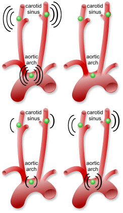
Specialized receptors in arteries monitor blood pressure and chemistry:
Baroreceptors: Detect changes in blood pressure (located in aortic arch and carotid arteries).
Chemoreceptors: Monitor pH, CO2, and O2 levels, important for respiratory and cardiovascular regulation.
Capillaries
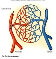
Capillaries are the exchange vessels, allowing passage of nutrients, wastes, and hormones between blood and tissues. Precapillary sphincters regulate blood flow into capillary beds, and metarterioles link arterioles to capillaries.
Types of Capillaries
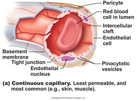
Continuous capillaries: Least permeable, found in skin and muscle; tight junctions and intercellular clefts allow passage of small solutes.
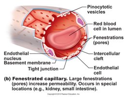
Fenestrated capillaries: Have pores (fenestrations) for rapid passage of molecules, found in kidneys and small intestine.
Sinusoidal (discontinuous) capillaries: Large gaps and fenestrations, allow passage of proteins and cells, found in liver, bone marrow, and spleen.
Veins
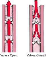
Veins are capacitance vessels, carrying blood back to the heart under lower pressure. They have thinner walls, larger lumens, and contain valves to prevent backflow. The skeletal muscle pump aids venous return, especially from the limbs.
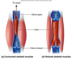
Venules: Smallest veins, receive blood from capillaries.
Medium veins: Contain valves, e.g., radial and saphenous veins.
Large veins: Include the venae cavae, which return blood to the heart.
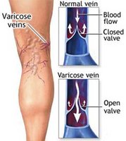
Clinical Note: Varicose Veins
Varicose veins occur when superficial veins become stretched and valves fail, often due to increased pressure from pregnancy, obesity, or prolonged standing. Hemorrhoids are a form of varicose veins in the anal canal.
Circulatory Routes
Types of Circulatory Pathways
Portal system: Blood flows through two consecutive capillary beds before returning to the heart (e.g., hepatic portal system).
Arteriovenous shunt: Artery flows directly into a vein, bypassing capillaries.
Venous anastomosis: Multiple veins drain the same tissue, providing alternate routes for blood return.
Arterial anastomosis: Multiple arteries supply the same region, providing collateral circulation.
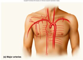
Major Arteries and Veins
The Aorta and Its Branches
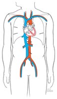
The aorta is the largest artery, distributing oxygen-rich blood to all systemic arteries. Major branches include the ascending aorta, aortic arch, thoracic aorta, and abdominal aorta.
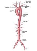
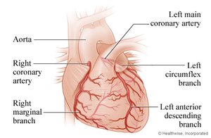
Coronary Arteries
The right and left coronary arteries branch from the ascending aorta to supply the heart muscle itself.
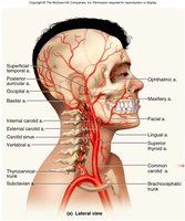
Arteries of the Head, Neck, and Limbs
Common carotid arteries: Branch into internal (brain) and external (face, scalp) carotids.
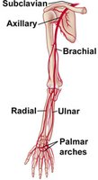
Subclavian artery: Supplies the upper limb, changing names as it passes through regions (axillary, brachial, radial, ulnar).
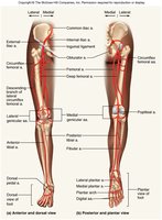
Femoral artery: Main artery of the lower limb, continuing as the popliteal and tibial arteries.
Major Veins
Superior and inferior vena cavae: Return deoxygenated blood from the upper and lower body to the right atrium.
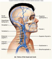
Brachiocephalic veins: Drain blood from the head, neck, and upper limbs into the superior vena cava.
Jugular veins: Internal jugular drains the brain; external jugular drains superficial head and neck structures.
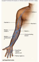
Great saphenous vein: Longest vein in the body, often used in coronary bypass surgery.
Hemodynamics: Blood Flow, Pressure, and Resistance
Principles of Blood Flow
Blood flow is determined by the pressure gradient and resistance within the vessels. Flow is directly proportional to the pressure difference and inversely proportional to resistance:
Perfusion: Rate of blood flow per given mass of tissue (ml/min/g).
Factors affecting flow: vessel diameter, blood viscosity, vessel length.
Blood Pressure
Blood pressure is the force exerted by blood against vessel walls, driving blood through the circulatory system. It is measured as systolic (during ventricular contraction) and diastolic (during relaxation) pressures, typically at the brachial artery.
Normal adult BP: <120/80 mm Hg
Hypertension: >130/80 mm Hg
Hypotension: Abnormally low BP, may result from blood loss or dehydration
Blood Pressure Changes in the Vascular Tree
Vessel | Pressure (mm Hg) |
|---|---|
Aorta | 120 |
Arteries | 120–40 |
Arterioles | 40–25 |
Capillaries | 25–12 |
Venules | 12–8 |
Veins | 10–5 |
Vena cavae | 2–0 |
Peripheral Resistance
Blood viscosity: Increased viscosity (e.g., polycythemia) raises resistance and BP; decreased viscosity (e.g., anemia) lowers BP.
Vessel length: Longer vessels increase resistance.
Vessel radius: Most adjustable; vasoconstriction increases resistance and BP, vasodilation decreases them.
Regulation of Blood Pressure and Flow
Local, Neural, and Hormonal Control
Local control: Vasoactive chemicals (e.g., histamine) and angiogenesis (new vessel growth) adjust local blood flow.
Neural control: The vasomotor center in the medulla oblongata integrates baroreflexes (pressure) and chemoreflexes (chemistry) to regulate vessel diameter.
Hormonal control: Hormones such as angiotensin II, ADH, atrial natriuretic factor, epinephrine, and norepinephrine influence blood volume and vessel tone.
Capillary Exchange Mechanisms
Modes of Exchange
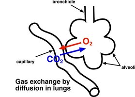
Diffusion: Movement of small molecules (e.g., O2, CO2, glucose) from high to low concentration.
Transcytosis: Transport of larger molecules (e.g., fatty acids, hormones) via vesicles across endothelial cells.
Filtration and reabsorption: Driven by hydrostatic and osmotic pressures; filtration occurs at the arterial end, reabsorption at the venous end.
Edema
Edema is the accumulation of excess fluid in tissues, caused by increased capillary filtration, reduced capillary reabsorption, or lymphatic obstruction. It can lead to tissue necrosis, pulmonary edema, or circulatory shock.
Venous Return and Circulatory Shock
Mechanisms of Venous Return
Pressure gradient from venules to the heart
Gravity (for blood from head and neck)
Skeletal muscle pump (in limbs)
Thoracic pump (during inhalation)
Cardiac suction (expanding atrial space)
Circulatory Shock
Cardiogenic shock: Inadequate heart pumping (e.g., myocardial infarction)
Low venous return (LVR) shock: Due to blood loss, venous pooling, or obstruction
Neurogenic shock: Sudden vasodilation from emotional stress or trauma
Clinical Correlations: TIAs and CVAs
Transient Ischemic Attacks (TIAs)
Brief episodes of cerebral ischemia causing dizziness, weakness, or loss of vision; often a warning sign of impending stroke.
Cerebrovascular Accidents (CVAs, Stroke)
Brain infarction due to ischemia from atherosclerosis, thrombosis, or ruptured aneurysm. Effects range from mild to fatal, including paralysis, loss of sensation, or speech impairment. Recovery depends on collateral circulation and neuronal plasticity.
