 Indietro
Chapter 21: The Immune System – Innate and Adaptive Body Defenses
Guida di studio - Note intelligenti
 Appunti personalizzati basati sui tuoi materiali, ampliati con definizioni chiave, esempi e contesto.
The Immune System: Overview
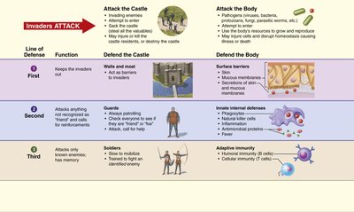
Lines of Defense
The immune system is a functional system that provides resistance to disease-causing microorganisms. It operates through three main lines of defense, each with distinct mechanisms and roles:
First Line: Surface barriers such as skin and mucous membranes act as physical and chemical barriers to prevent pathogen entry.
Second Line: Innate internal defenses include phagocytes, natural killer cells, antimicrobial proteins, inflammation, and fever. These mechanisms respond rapidly to threats that breach the first line.
Third Line: Adaptive (specific) defenses involve lymphocytes (B and T cells) and provide targeted responses to specific pathogens, including memory for future encounters.
First Line of Defense: Surface Barriers
Skin and Mucous Membranes
Surface barriers are the body's initial protection against pathogens. They include:
Intact skin epidermis: Forms a mechanical barrier.
Acid mantle of skin: Skin secretions create an acidic environment, inhibiting bacterial growth.
Keratin: Provides resistance against acids, alkalis, and bacterial enzymes.
Mucous membranes: Line body cavities and trap pathogens.
Mucus, nasal hairs, cilia: Trap and propel debris-laden mucus away from respiratory passages.
Gastric juice: Contains hydrochloric acid and enzymes that destroy pathogens.
Lacrimal secretion (tears), saliva, urine: Cleanse and contain lysozyme, an enzyme that destroys microorganisms.
Second Line of Defense: Innate Internal Defenses
Phagocytes
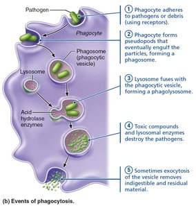
Phagocytes are cells that engulf and destroy pathogens. The process of phagocytosis involves several steps:
Adherence: Phagocyte adheres to pathogens or debris.
Engulfment: Forms pseudopods to engulf particles, creating a phagosome.
Fusion: Lysosome fuses with phagosome, forming a phagolysosome.
Destruction: Toxic compounds and lysosomal enzymes destroy pathogens.
Exocytosis: Indigestible material is expelled.
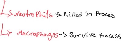
Neutrophils are killed in the process, while macrophages survive and can continue to phagocytose.
Natural Killer (NK) Cells
NK cells are large granular lymphocytes that patrol blood and lymph, targeting cells with abnormal or missing "self" proteins (MHC). They kill cancer and virus-infected cells before the adaptive immune system is activated and secrete chemicals to enhance inflammation.
Inflammation
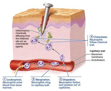
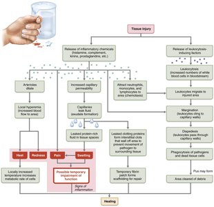
Inflammation is a nonspecific response to tissue injury, caused by trauma, heat, chemicals, or infection. It prevents pathogen spread, disposes of debris, alerts the adaptive immune system, and sets the stage for repair. The stages include:
Inflammatory Chemical Release: Histamine, kinins, prostaglandins, complement, and cytokines are released, causing vasodilation and increased permeability.
Vasodilation and Increased Vascular Permeability: Local hyperemia brings more immune cells; exudate causes swelling and pain.
Phagocyte Mobilization: Leukocytes migrate to the site of injury.
Antimicrobial Proteins
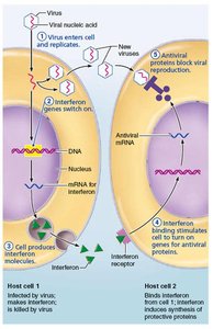
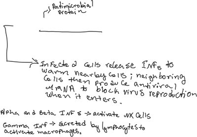
Antimicrobial proteins include interferons and complement proteins. Interferons are released by infected cells to warn nearby cells, which then produce antiviral mRNA to block virus reproduction. Alpha and beta interferons activate NK cells, while gamma interferon activates macrophages.
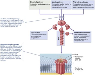
Complement proteins enhance inflammation, opsonize pathogens, and form membrane attack complexes (MAC) to lyse target cells.
Fever
Fever is a systemic response to infection. Pyrogens released by leukocytes and macrophages act on the hypothalamus to raise body temperature, enhancing immune response and inhibiting bacterial growth.
Third Line of Defense: Adaptive (Specific) Defenses
Key Differences Between Innate and Adaptive Defenses
Feature | Innate Defenses | Adaptive Defenses |
|---|---|---|
Cells Involved | Diverse cells (NK, phagocytes, etc.) | B and T lymphocytes |
Specificity | Nonspecific | Specific (recognize antigens) |
Response Time | Fast | Slow |
Memory | None | Has memory |
Scope | Local | Systemic |
Antigens
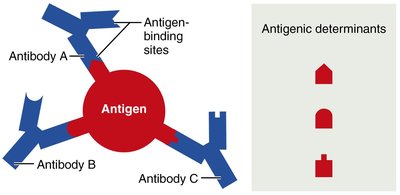
Antigens are substances that trigger adaptive defenses. Complete antigens have immunogenicity and reactivity. Incomplete antigens (haptens) become immunogenic when attached to protein carriers. Most antigens have several antigenic determinants, which are the specific parts recognized by antibodies or lymphocyte receptors.
Self-Antigens and MHC Proteins
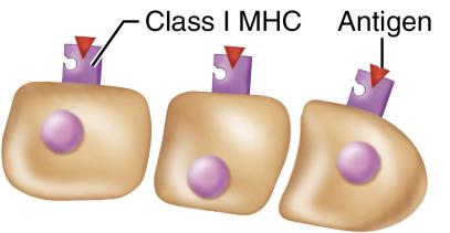
Self-antigens are proteins on cell surfaces that are not antigenic to self but are to others. Major Histocompatibility Complex (MHC) proteins are glycoproteins that present antigens to T cells:
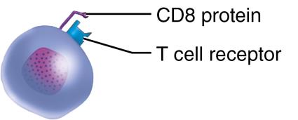
Class I MHC: Displayed by all nucleated cells except RBCs; recognized by CD8 cells and cytotoxic T cells.
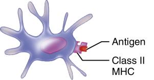
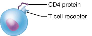
Class II MHC: Displayed by antigen-presenting cells (APCs); recognized by CD4 cells and helper T cells.
Cells of the Adaptive Immune Response
The adaptive immune system involves:
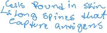
Antigen-presenting cells (APCs): Dendritic cells, macrophages, and B cells that present antigens to T cells.
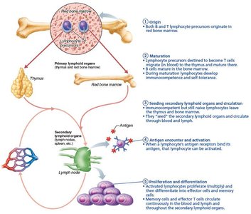
B lymphocytes: Provide humoral immunity by producing antibodies.
T lymphocytes: Provide cellular immunity by directly attacking infected or abnormal cells.
B and T Lymphocytes: Comparison
Feature | B Lymphocytes | T Lymphocytes |
|---|---|---|
Type of Response | Humoral | Cellular |
Antibody Secretion | Yes | No |
Primary Targets | Extracellular pathogens | Intracellular pathogens, cancer cells |
Site of Origin | Red bone marrow | Red bone marrow |
Site of Maturation | Red bone marrow | Thymus |
Effector Cells | Plasma cells | Cytotoxic, helper, regulatory T cells |
Memory Cell Formation | Yes | Yes |
Clonal Selection of B Cells
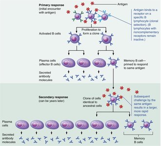
When a B cell encounters its specific antigen, it undergoes clonal selection, proliferating to form a clone of plasma cells (which secrete antibodies) and memory B cells (which respond to future encounters).
Antibody Structure and Classes
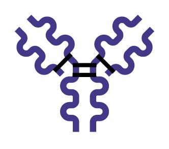
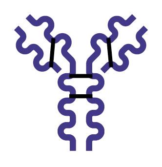
Antibodies (immunoglobulins) are Y-shaped proteins composed of two heavy and two light chains, with variable and constant regions. The variable region binds antigens, while the constant region determines the antibody class and function.
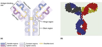
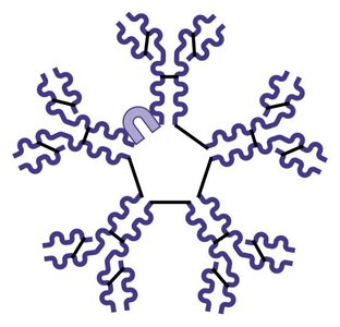
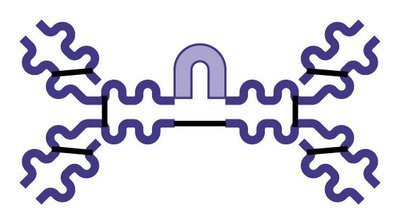
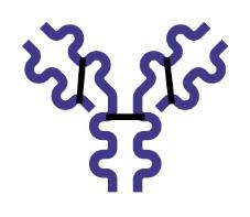
There are five major classes of immunoglobulins:
IgM: First antibody secreted; potent agglutinating agent; activates complement.
IgA: Found in body secretions; prevents pathogen attachment to epithelial surfaces.
IgD: Functions as a B cell antigen receptor.
IgG: Most abundant; main antibody of secondary responses; crosses placenta.
IgE: Binds to mast cells and basophils; triggers allergic reactions and inflammation.
Mechanisms of Antibody Action
Antibodies act by:
Neutralization: Blocking harmful effects of toxins or pathogens.
Agglutination: Clumping antigens together.
Precipitation: Causing soluble antigens to settle out.
Complement activation: Triggering the complement cascade to lyse pathogens.
Primary and Secondary Humoral Responses
The primary response occurs upon first exposure to an antigen, producing plasma cells and memory B cells. The secondary response is faster and stronger due to memory cells.
Active and Passive Humoral Immunity
Immunity can be:
Active: Produced by exposure to antigen (natural infection or vaccination).
Passive: Acquired by receiving antibodies (maternal antibodies or injection).
Immune System Homeostatic Imbalances
Immunodeficiency
Immunodeficiency is a condition where immune function is impaired, either congenitally or acquired, affecting cells or molecules like complement or antibodies.
Autoimmune Diseases
Autoimmune diseases occur when the immune system fails to distinguish self from foreign antigens, often due to self-reactive lymphocytes or molecular mimicry.
Hypersensitivities
Hypersensitivities are immune responses to harmless threats that cause tissue damage. Types include:
Immediate (Type I): Allergies mediated by IgE; rapid onset.
Subacute (Type II and III): Mediated by IgM and IgG; slower onset, longer duration.
Delayed (Type IV): Mediated by T cells; takes 1–3 days to appear.
Summary Table: Immunoglobulin Classes
Class | Structure | Key Characteristics |
|---|---|---|
IgM | Pentamer | First secreted, activates complement, potent agglutinating agent |
IgA | Dimer | Found in secretions, prevents pathogen attachment |
IgD | Monomer | B cell antigen receptor |
IgG | Monomer | Most abundant, crosses placenta, activates complement |
IgE | Monomer | Triggers allergic reactions, binds mast cells/basophils |
Additional info:
Pattern recognition receptors (e.g., Toll-like receptors) are crucial for innate immune responses.
Helper T cells coordinate both humoral and cellular responses.
Organ transplants require immunosuppressive therapy to prevent rejection, increasing susceptibility to infection.
