 Indietro
Chapter 23: The Digestive System – Anatomy & Physiology Study Notes
Guida di studio - Note intelligenti
 Appunti personalizzati basati sui tuoi materiali, ampliati con definizioni chiave, esempi e contesto.
The Digestive System
Overview and Functions
The digestive system is responsible for processing food, breaking it down into nutrients, absorbing these nutrients into the bloodstream, and eliminating indigestible remains. It consists of a series of organs and accessory structures that work together to accomplish these tasks.
Ingestion: Taking in food through the mouth.
Mechanical Breakdown: Chewing, mixing with saliva, and churning in the stomach and intestines.
Digestion: Enzymatic breakdown of complex food molecules into simpler chemical building blocks.
Absorption: Movement of digested fragments from the GI tract lumen into blood or lymph.
Defecation: Elimination of indigestible substances via the anus.
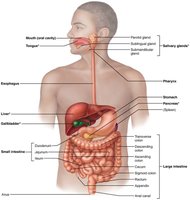
Organs of the Digestive System
The organs are divided into two groups: the alimentary canal and accessory digestive organs.
Alimentary Canal (GI Tract): Continuous muscular tube from mouth to anus. Includes: mouth, pharynx, esophagus, stomach, small intestine, large intestine, rectum, and anus.
Accessory Digestive Organs: Teeth, tongue, gallbladder, salivary glands, liver, and pancreas. These produce secretions that aid in digestion.
Digestive Processes
Six essential activities are involved in food processing:
Ingestion
Propulsion: Movement of food via swallowing and peristalsis (alternating waves of contraction and relaxation).
Mechanical Breakdown
Digestion
Absorption
Defecation
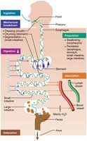
Organization and Anatomy of the Digestive System
Peritoneum and Mesenteries
The peritoneum is a serous membrane lining the abdominal cavity, consisting of:
Visceral Peritoneum: Covers external surfaces of most digestive organs.
Parietal Peritoneum: Lines the body wall.
Peritoneal Cavity: Fluid-filled space between the two peritoneums, lubricating mobile organs.
Mesentery: Double layer of peritoneum that provides routes for blood vessels, lymphatics, and nerves, holds organs in place, and stores fat.
Intraperitoneal Organs: Located within the peritoneum.
Retroperitoneal Organs: Located outside or posterior to the peritoneum (e.g., most of pancreas, duodenum, parts of large intestine).
Blood Supply: Splanchnic Circulation
Splanchnic Circulation: Includes arteries branching off the aorta (hepatic, splenic, left gastric, inferior and superior mesenteric arteries) and hepatic portal circulation (drains nutrient-rich blood from digestive organs to the liver).
Control of Digestive System
Enteric Nervous System
The GI tract has its own nervous system, the enteric nervous system (gut brain), which contains more neurons than the spinal cord. Enteric neurons communicate extensively and control motility and secretions.
Regulation: Digestive activity is provoked by mechanical and chemical stimuli (stretch, osmolarity, pH, presence of substrates).
Effectors: Smooth muscle and glands respond to stimuli, mixing and moving contents, and secreting digestive juices or hormones.
Functional Anatomy of the Digestive System
Mouth and Associated Organs
The mouth (oral cavity) is where food is chewed and mixed with saliva, initiating digestion and swallowing. It is bounded by lips, cheeks, palate, and tongue.
Palate: Roof of the mouth, divided into hard palate (bone) and soft palate (muscle). The uvula is a fingerlike projection from the soft palate.
Tongue: Composed of skeletal muscle, grips and mixes food, forms the bolus, initiates swallowing, speech, and taste. The lingual frenulum attaches the tongue to the floor of the mouth.
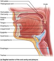
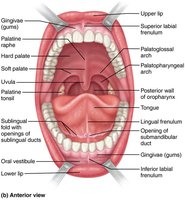
Salivary Glands
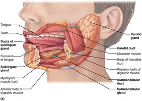
Salivary glands produce saliva, which cleanses the mouth, dissolves food chemicals, moistens food, compacts it into a bolus, and begins starch breakdown with amylase.
Major Salivary Glands: Parotid (anterior to ear), Submandibular (medial to mandible), Sublingual (under tongue).
Saliva: Contains enzymes, electrolytes, and mucins.
Teeth and Dentition
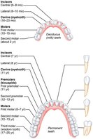
Teeth are located in sockets in the mandible and maxilla. They are essential for mastication (chewing), which tears and grinds food.
Primary Dentition: 20 deciduous (milk) teeth erupt between 6–24 months.
Permanent Dentition: 32 teeth, including incisors (cutting), canines (tearing), premolars (grinding/crushing), and molars (grinding).
Dental Formula: Describes the number and types of teeth in each quadrant.
Tooth and Gum Disease
Dental caries (cavities) result from demineralization of enamel and dentin by bacterial action. Dental plaque adheres to teeth, and acid from bacteria dissolves calcium salts. Gingivitis and periodontitis are gum diseases caused by plaque and tartar buildup, leading to infection and potential systemic effects.
Prevention: Daily flossing and brushing.
Risk Factors: Smoking, diabetes, oral piercings.
Systemic Effects: Periodontitis may increase risk of heart disease and stroke.
Clinical Homeostatic Imbalances
Peritonitis: Inflammation of the peritoneum, potentially life-threatening.
Ankyloglossia: Congenital short lingual frenulum, restricting tongue movement.
Xerostomia: Dry mouth due to insufficient saliva.
Impacted Tooth: Tooth trapped in jawbone, often wisdom teeth.
Summary Table: Types of Teeth and Their Functions
Type of Tooth | Shape | Function |
|---|---|---|
Incisors | Chisel-shaped | Cutting |
Canines | Fanglike | Tearing/Piercing |
Premolars | Broad crowns with rounded cusps | Grinding/Crushing |
Molars | Broad crowns, rounded cusps | Grinding |
Key Equations
Dental formula for adult human:
(per quadrant)
Total teeth:
Additional info:
Clinical notes and disease prevention strategies are expanded for academic completeness.
Table is inferred from classification of teeth in the notes.
