 Indietro
Chapter 24: The Urinary System – Structure and Function
Guida di studio - Note intelligenti
 Appunti personalizzati basati sui tuoi materiali, ampliati con definizioni chiave, esempi e contesto.
Urinary System Overview
Major Organs and Functions
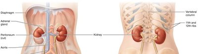
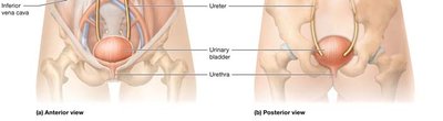
The urinary system is essential for maintaining homeostasis by filtering blood, removing metabolic wastes, and regulating fluid, electrolyte, and acid-base balance. It consists of the paired kidneys and the urinary tract, which includes the ureters, urinary bladder, and urethra.
Kidneys: Filter blood, remove waste, regulate blood pressure, and produce hormones.
Urinary Tract: Transports, stores, and eliminates urine.
Adrenal Glands: Sit atop each kidney and produce hormones like aldosterone.
Anatomy of the Kidneys
External Structure and Location
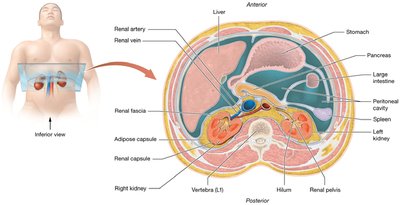
The kidneys are retroperitoneal organs, partially protected by the 11th and 12th ribs. The right kidney is slightly lower than the left due to the liver's position. Each kidney is covered by three layers of connective tissue:
Renal Fascia: Anchors the kidney to surrounding structures.
Adipose Capsule: Cushions and protects against physical shock.
Renal Capsule: Protects from infection and trauma.
External and Internal Features
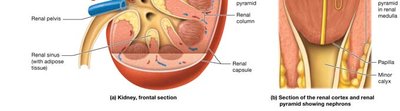
The kidney's hilum is the entry and exit point for blood vessels, nerves, lymphatics, and the ureter. The renal sinus is a cavity containing connective tissue and the renal pelvis, which collects urine.
Internal Anatomy: Cortex and Medulla
The kidney is divided into the renal cortex and renal medulla:
Renal Cortex: Contains most blood vessels and the renal corpuscles of nephrons.
Renal Medulla: Contains renal pyramids and tubules, separated by renal columns.
Renal Papilla: Apex of pyramid, drains urine into minor calyx.
Microscopic Anatomy: Nephrons and Collecting System
Nephron Structure
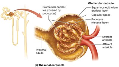
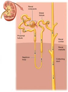
Nephrons are the functional units of the kidney, each composed of a renal corpuscle and a renal tubule. The renal corpuscle consists of:
Glomerulus: Fenestrated capillaries for filtration.
Glomerular Capsule (Bowman's Capsule): Double-walled structure with parietal (outer) and visceral (inner, podocytes) layers.
Capsular Space: Between parietal and visceral layers, where filtrate collects.
Renal Tubule and Collecting System
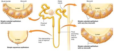
The renal tubule transports filtrate and allows reabsorption and secretion:
Proximal Tubule
Nephron Loop (Loop of Henle): Descending and ascending limbs
Distal Tubule
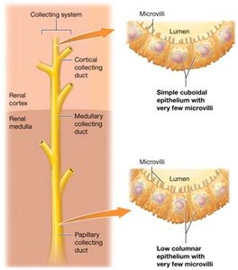
Collecting Duct: Drains into papillary duct, then calyces
Juxtaglomerular Apparatus (JGA)
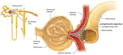
The JGA is located between the ascending limb of the nephron loop and the afferent/efferent arterioles. It consists of macula densa and juxtaglomerular cells, regulating filtrate flow, blood pressure, and urine volume.
Types of Nephrons
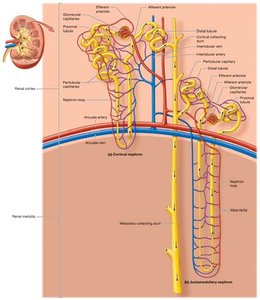
Cortical Nephrons: 80% of nephrons, short loops, mainly in cortex.
Juxtamedullary Nephrons: Long loops, deep cortex near medulla, surrounded by vasa recta, responsible for concentrated urine.
Blood Supply of the Kidneys
Pathway of Blood Flow
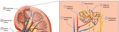
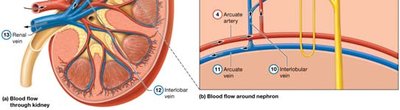
Blood enters the kidney via the renal artery and follows a branching pathway:
Renal artery → Segmental artery → Interlobar artery → Arcuate artery → Interlobular (cortical radiate) artery → Afferent arteriole → Glomerulus → Efferent arteriole → Peritubular capillaries → Interlobular vein → Arcuate vein → Interlobar vein → Renal vein
Renal Physiology: Urine Formation
Three Mechanisms of Urine Formation
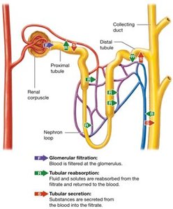
Glomerular Filtration: Movement of water and solutes from blood into filtrate at the renal corpuscle.
Tubular Reabsorption: Essential materials and water move from filtrate back into blood at the renal tubule.
Tubular Secretion: Removal of waste and H+ from blood into tubule.
Filtration Membrane
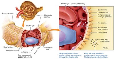
The filtration membrane consists of three layers:
Fenestrated glomerular capillary endothelial cells: Prevent passage of formed elements.
Basal lamina: Collagen fibers prevent passage of large proteins.
Visceral layer (podocytes): Filtration slits allow only small molecules to pass.
Glomerular Filtration Rate (GFR)
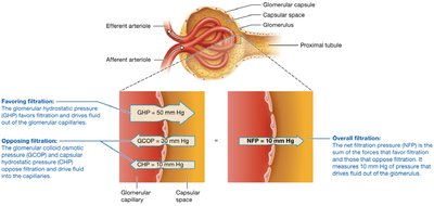
GFR is the amount of filtrate produced per minute (normal: 120–125 ml/min). It is determined by hydrostatic and osmotic pressures:
Glomerular hydrostatic pressure (GHP): Promotes filtration (≈50 mm Hg).
Glomerular colloid osmotic pressure (GCOP): Promotes reabsorption (≈30 mm Hg).
Capsular hydrostatic pressure (CHP): Promotes reabsorption (≈10 mm Hg).
Net Filtration Pressure (NFP):
The net filtration pressure is calculated as:
Regulation of GFR
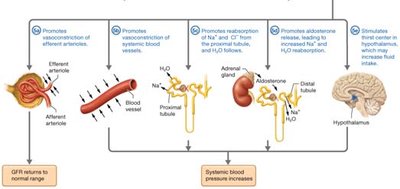
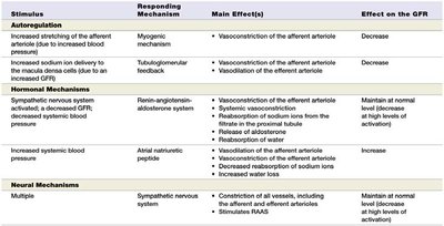
Autoregulation: Myogenic mechanism and tubuloglomerular feedback maintain GFR within normal range.
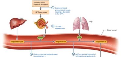
Hormonal Regulation: Renin-angiotensin-aldosterone system (RAAS) and atrial natriuretic peptide (ANP) adjust GFR and blood pressure.
Neural Regulation: Sympathetic nervous system influences GFR during stress.
Stimulus | Responding Mechanism | Main Effect(s) | Effect on the GFR |
|---|---|---|---|
Increased stretching of afferent arteriole | Myogenic mechanism | Vasoconstriction of afferent arteriole | Decrease |
Increased sodium ion delivery to macula densa | Tubuloglomerular feedback | Vasoconstriction of afferent arteriole, vasodilation of efferent arteriole | Decrease |
Sympathetic nervous system activation | Renin-angiotensin-aldosterone system | Vasoconstriction, sodium reabsorption, aldosterone release | Maintain at normal level |
Increased systemic blood pressure | Atrial natriuretic peptide | Dilation of afferent arteriole, constriction of efferent arteriole | Increase |
Multiple | Sympathetic nervous system | Constriction of vessels, stimulate RAAS | Maintain at normal level |
Tubular Reabsorption and Secretion
Principles and Routes
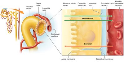
Reabsorption moves substances from filtrate to blood; secretion moves substances from blood to filtrate. Two routes:
Paracellular Route: Passive movement between cells.
Transcellular Route: Active movement through cells, often requiring carrier proteins.
Carrier-Mediated Transport
Facilitated Diffusion: Passive transport with concentration gradient.
Primary Active Transport: Uses ATP to move solutes against gradient.
Secondary Active Transport: Uses gradient from primary transport to move other solutes.
Reabsorption in the Proximal Tubule
65% of electrolytes
Nearly 100% of nutrients
90% of bicarbonate ions
65% of water
Secretion involves hydrogen ions, nitrogenous wastes, and drugs.
Sodium and Organic Solute Reabsorption
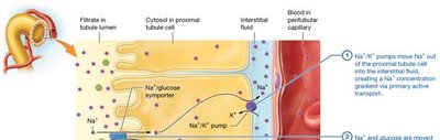
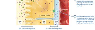
Sodium is reabsorbed via leak channels, symporters, and antiporters. Glucose and other solutes are reabsorbed using sodium gradients.
Bicarbonate Ion Reabsorption
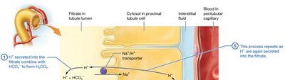
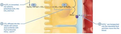
Bicarbonate is reabsorbed by combining with H+ to form CO2 and H2O, which are then converted back to HCO3- inside the cell and transported to blood.
Obligatory Water Reabsorption
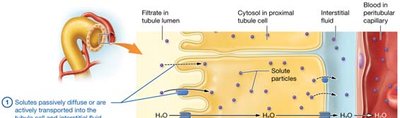
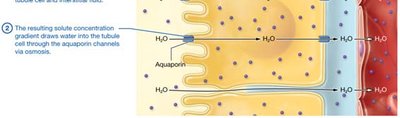
Water follows solutes by osmosis through aquaporin channels, driven by solute concentration gradients.
Summary
This chapter provides a comprehensive overview of the urinary system, focusing on the anatomy and physiology of the kidneys, nephron structure, blood supply, and the mechanisms of urine formation, reabsorption, and secretion. Understanding these processes is essential for grasping how the body maintains homeostasis and eliminates waste.
