Indietro
Chapter 24: The Urinary System – Structure and Function
Guida di studio - Note intelligenti
 Appunti personalizzati basati sui tuoi materiali, ampliati con definizioni chiave, esempi e contesto.
Urinary System Overview
Main Components and Functions
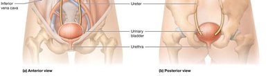
The urinary system is essential for maintaining homeostasis by filtering blood, removing waste, and regulating fluid, electrolyte, and acid-base balance. It consists of the paired kidneys and the urinary tract, which includes the ureters, urinary bladder, and urethra.
Kidneys: Filter blood, remove metabolic wastes, regulate blood pressure, and maintain fluid and electrolyte balance.
Urinary Tract: Transports, stores, and eliminates urine.
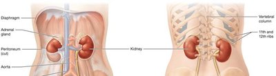
Adrenal Glands: Sit atop each kidney and are involved in hormone production.
Anatomy of the Kidneys
External Structure
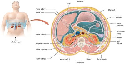
The kidneys are retroperitoneal organs, partially protected by the 11th and 12th ribs. The right kidney is slightly lower than the left due to the liver. Each kidney is covered by three layers:
Renal Fascia: Anchors the kidney to surrounding structures.
Adipose Capsule: Cushions and protects the kidney from physical shock.
Renal Capsule: Protects from infection and trauma.
Internal Structure
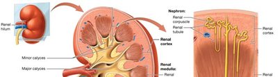
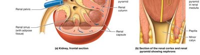
The kidney's internal anatomy includes the renal cortex, renal medulla, and renal pelvis. The cortex contains most blood vessels and the renal corpuscles of nephrons. The medulla consists of renal pyramids separated by renal columns, and the renal pelvis collects urine before it enters the ureter.
Renal Cortex: Houses renal corpuscles and blood vessels.
Renal Medulla: Contains renal pyramids and tubules.
Renal Pelvis: Central cavity for urine collection.
Microscopic Anatomy: Nephrons
Nephron Structure and Function
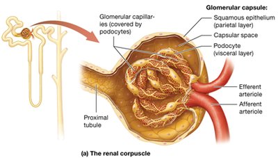
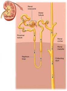
Nephrons are the functional units of the kidney, responsible for urine formation. Each nephron consists of a renal corpuscle and a renal tubule. The renal corpuscle includes the glomerulus and Bowman's capsule, where filtration occurs. The renal tubule processes filtrate through reabsorption and secretion.
Renal Corpuscle: Glomerulus (fenestrated capillaries) and Bowman's capsule (parietal and visceral layers).
Renal Tubule: Proximal tubule, nephron loop (descending and ascending limbs), distal tubule, and collecting duct.
Histology of Renal Tubule
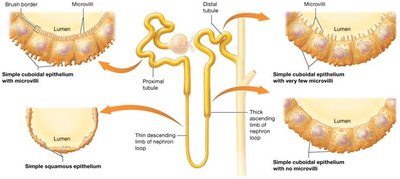
The renal tubule is lined with different types of epithelial cells, each adapted for specific functions such as absorption and secretion.
Proximal Tubule: Simple cuboidal epithelium with microvilli for increased absorption.
Nephron Loop: Thin descending limb (simple squamous), thick ascending limb (cuboidal with few microvilli).
Distal Tubule: Cuboidal with very few microvilli.
Juxtaglomerular Apparatus (JGA)
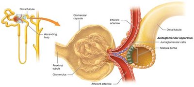
The JGA is a specialized structure that regulates blood flow and filtration rate. It consists of macula densa cells and juxtaglomerular cells, located between the ascending limb of the nephron loop and the afferent/efferent arterioles.
Function: Regulates urine volume, blood volume, and blood pressure.
Blood Supply of the Kidneys
Pathway of Blood Flow
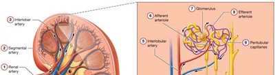
Blood enters the kidney via the renal artery and passes through a series of vessels before being filtered in the glomerulus. The pathway is:
Renal artery
Segmental artery
Interlobar artery
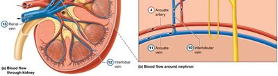
Arcuate artery
Interlobular (cortical radiate) artery
Afferent arteriole
Glomerulus
Efferent arteriole
Peritubular capillaries
Interlobular vein
Arcuate vein
Interlobar vein
Renal vein