 Indietro
Chapter 3: The Cell – Structure, Function, and Microscopy
Guida di studio - Note intelligenti
 Appunti personalizzati basati sui tuoi materiali, ampliati con definizioni chiave, esempi e contesto.
Chapter 3: The Cell
Introduction to Cell Theory
The cell is the fundamental unit of life, and cell theory forms the basis of modern biology. The development of microscopes enabled scientists to observe cells and led to the formulation of cell theory.
Cell Theory: All living things are composed of one or more cells; the cell is the basic unit of life; all new cells arise from existing cells.
Historical Context: Robert Hooke coined the term "cell" in 1665. Schleiden and Schwann unified cell theory in the 1830s.
Common Components of All Cells
Despite diversity, all cells share four essential components that enable their function and survival.
Plasma Membrane: A phospholipid bilayer that separates the cell's interior from its environment.
Cytoplasm: Jelly-like substance containing fluid, proteins, and organelles.
DNA: Genetic material responsible for heredity and cellular function.
Ribosomes: Structures that synthesize proteins.
Cell Size and Surface Area-to-Volume Ratio
Cell size is measured in micrometers (µm), and cells must remain small to efficiently exchange materials with their environment.
Average Human Cell: Approximately 10 µm in diameter.
Surface Area-to-Volume Ratio: Smaller cells have a higher ratio, facilitating nutrient uptake and waste removal.
Importance: As cells grow larger, their surface area does not increase as quickly as their volume, limiting efficient exchange.
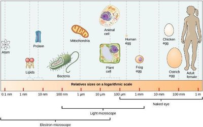
Figure: Relative sizes of atoms, molecules, cells, and organisms, showing the scale at which different microscopes are used.
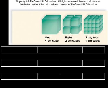
Figure: Surface area-to-volume ratio illustrated with cubes of different sizes. Smaller cubes have more surface area relative to their volume.
Microscopy
Types of Microscopes
Microscopes are essential tools for visualizing cells and their components. Different types of microscopes offer varying levels of magnification and resolution.
Compound Light Microscope: Uses glass lenses and light to magnify specimens up to 400–1,000 times. Orientation of the image is reversed.
Dissecting Microscope: Lower magnification, used for larger objects and tissues. Produces upright images.
Electron Microscope: Uses electron beams for much higher magnification and resolution. Includes transmission (2-D, internal structures) and scanning (3-D, surface details) types.
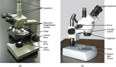
Figure: Anatomy of a compound light microscope (a) and a dissecting microscope (b).
Magnification and Resolving Power
Magnification is the enlargement of an object, while resolving power is the ability to distinguish adjacent structures.
Light Microscope: Magnifies up to 1,000×; resolving power ~0.1 µm.
Transmission Electron Microscope: Magnifies up to 100,000×; resolving power ~0.01 µm.
Human Eye: Resolving power ~100 µm.
Microscopy Images
Different microscopes produce distinct images, revealing various aspects of cell structure.
Light Micrograph: Shows stained cells and tissues.
Transmission Electron Micrograph: Reveals internal cell structures in detail.
Scanning Electron Micrograph: Provides 3-D views of cell surfaces.
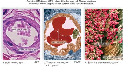
Figure: Comparison of micrographs produced by different types of microscopes.
Prokaryotic vs. Eukaryotic Cells
Prokaryotic Cells
Prokaryotic cells are simpler and evolved first. They lack membrane-bound organelles and a nucleus.
Organisms: Bacteria and Archaea.
Cell Wall: Made of peptidoglycan; provides protection and shape.
Capsule: Polysaccharide layer for attachment and protection.
Flagella: Used for locomotion.
Pili: Used for genetic exchange (conjugation).
DNA: Located in the nucleoid region.
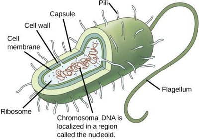
Figure: Generalized structure of a prokaryotic cell, showing key components.
Eukaryotic Cells and Endosymbiosis
Eukaryotic cells are more complex, containing membrane-bound organelles and a true nucleus. The endosymbiosis theory explains the origin of certain organelles.
Eukaryotic: Means "true nucleus"; includes animals, plants, fungi, and protists.
Organelles: Specialized structures such as mitochondria and chloroplasts.
Endosymbiosis: Theory that mitochondria and chloroplasts originated from prokaryotic cells engulfed by ancestral eukaryotes.
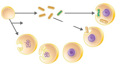
Figure: Model of endosymbiosis showing the origin of mitochondria and chloroplasts in eukaryotic cells.
Eukaryotic Cell Structure
Overview of Eukaryotic Cell Components
Eukaryotic cells contain numerous organelles, each with specialized functions necessary for cellular life.
Nucleus: Contains DNA and controls cellular activities.
Endoplasmic Reticulum (ER): Rough ER synthesizes proteins; smooth ER synthesizes lipids.
Golgi Apparatus: Modifies, sorts, and packages proteins and lipids.
Mitochondria: Site of cellular respiration and ATP production.
Lysosomes: Digest cellular waste.
Peroxisomes: Break down fatty acids and detoxify harmful substances.
Cytoskeleton: Maintains cell shape and enables movement.
Microvilli: Increase surface area for absorption.
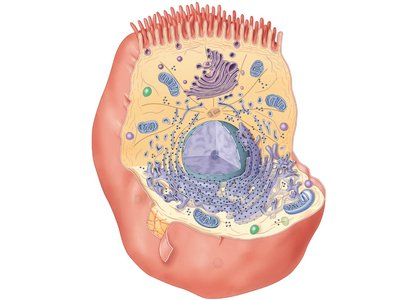
Figure: Eukaryotic cell showing major organelles and structures.
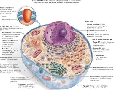
Figure: Detailed labeled diagram of a eukaryotic cell, highlighting the plasma membrane, cytoskeleton, nucleus, endoplasmic reticulum, mitochondria, and other organelles.
Summary Table: Resolving Power of the Eye and Common Microscopes
This table compares the magnification and resolving power of the human eye, light microscope, and transmission electron microscope.
Instrument | Magnification | Resolving Power |
|---|---|---|
Eye | N/A | 0.1 mm (100 μm) |
Light microscope | 1,000× | 0.0001 mm (0.1 μm) |
Transmission electron microscope | 100,000× (or greater) | 0.000001 mm (0.01 μm) |
Key Equations
Cell size and surface area-to-volume ratio are important for understanding cell function.
Micrometer:
Surface Area of a Cube:
Volume of a Cube:
Surface Area-to-Volume Ratio:
Understanding these ratios helps explain why cells are small and how they efficiently exchange materials with their environment.
