 Indietro
Chapter 4: Histology – The Study of Tissues
Guida di studio - Note intelligenti
 Appunti personalizzati basati sui tuoi materiali, ampliati con definizioni chiave, esempi e contesto.
Histology: The Study of Tissues
Introduction to Tissues
Histology is the branch of anatomy that studies tissues, which are groups of similar cells and their extracellular matrix working together to perform specific functions. All tissues are composed of a population of related cells and an extracellular matrix (ECM).
Tissue: Groups of similar cells and their external environment (ECM) working together for common functions.
Extracellular Matrix (ECM): Substances surrounding cells in tissue, providing structural and biochemical support.
Types of Tissue
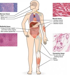
There are four primary tissue types in the human body, each with distinct functions and characteristics:
Epithelial Tissue: Tightly packed cells that line body surfaces and cavities, forming parts of glands; little ECM.
Connective Tissue: Connects all other tissues in the body; abundant ECM.
Muscle Tissue: Composed of cells that contract and generate force; little ECM.
Nervous Tissue: Made of cells that generate, send, and receive messages; variable ECM.
Extracellular Matrix (ECM)
Components of the ECM
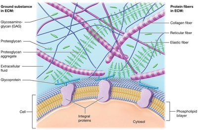
The ECM is a complex network of substances that surround cells in tissues. It provides strength, directs cell behavior, regulates development, mitosis, and survival, and holds cells in position.
Ground Substance: Contains water, ions, nutrients, and three types of macromolecules:
Glycosaminoglycans (GAGs): Negatively charged polysaccharide chains that attract positive ions and water, helping ECM resist compression.
Proteoglycans: GAGs bound to a protein core, forming aggregates that make ECM more solid and act as barriers to diffusion.
Glycoproteins: Cell-adhesion molecules (CAMs) that bind cell surface proteins and fibers, maintaining tissue architecture.
Protein Fibers: Provide tensile strength and structural support:
Collagen fibers: Resist tension and pressure.
Elastic fibers: Allow stretch and return to original shape.
Reticular fibers: Form scaffolding to support cells and ground substance.
Cell Junctions
Types of Cell Junctions
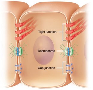
Cell junctions are specialized structures that link neighboring cells in tissues via integral proteins. There are three main types:
Tight (Occluding) Junctions: Integral proteins interlock to form a "zipper," preventing macromolecules from moving between cells. Some are "leaky." Found in blood vessels.
Desmosomes: Proteins lock together like buttons, allowing small spaces for material passage and increasing resistance to mechanical stress. Found in skin.
Gap Junctions: Channel proteins form small pores, allowing material to freely move between cells and enabling cell-to-cell communication, especially in cardiac muscle cells.
Epithelial Tissue
Functions and Components
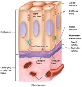
Epithelial tissue covers external body surfaces and lines body cavities, acting as a barrier. It is avascular and anchored to underlying connective tissue via the basement membrane.
Protection: Rapid mitosis for repair.
Immune Defense: Prevents microorganism entry.
Secretion: Forms glands producing secretions.
Transportation: Selectively permeable barriers.
Sensation: Houses sensory receptors.
Classification of Epithelia
Epithelia are classified by arrangement and shape of cells:
Arrangement:
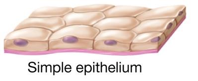
Simple: One layer of cells.
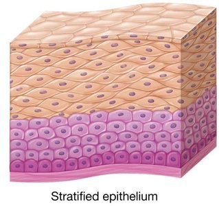
Stratified: Two or more layers.
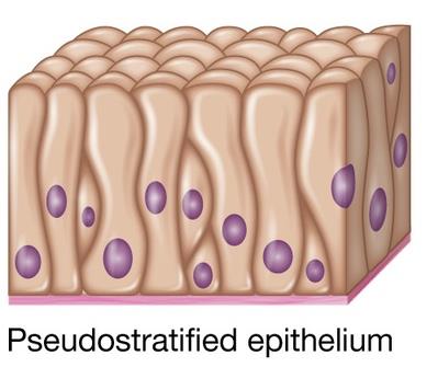
Pseudostratified: Single layer appearing multilayered.
Shape:
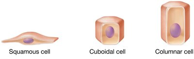
Squamous: Scalelike.
Cuboidal: Cubelike.
Columnar: Column-shaped.
Simple Epithelia
Simple epithelia consist of a single cell layer, ideal for diffusion and transport.
Simple Squamous Epithelium: Flat cells; diffusion; air sacs of lungs, lining blood vessels.
Simple Cuboidal Epithelium: Cube-shaped; diffusion and secretion; kidney tubules, thyroid gland.
Simple Columnar Epithelium: Column-shaped; microvilli, cilia, goblet cells; diffusion and secretion; small intestine, uterine tube.
Pseudostratified Columnar Epithelium: Appears stratified; ciliated, goblet cells; protection; respiratory passages.
Stratified Epithelia
Stratified epithelia have multiple cell layers, providing protection in areas of high stress.
Stratified Squamous Epithelia: Flat cells; protection; mouth, esophagus, vagina.
Stratified Cuboidal Epithelia: Cube-shaped; some absorption; ducts of sweat glands.
Stratified Columnar Epithelia: Rectangular cells; protection and absorption; ducts of salivary glands.
Transitional Epithelia: Cube and squamous cells; allows stretching; urinary bladder.
Glandular Epithelia
Types of Glands
Glands are structures that make and secrete products, formed from invaginated epithelial tissue. Products are released in two ways:
Exocrine Glands: Release secretions through a duct onto the apical surface; local actions.
Endocrine Glands: Release secretions directly into blood; lacks ducts; widespread actions.
Connective Tissue
Functions and Classification
Connective tissue binds, supports, protects, and transports materials throughout the body. It is classified as:
Connective Tissue Proper: Widely distributed; includes loose, dense, reticular, and adipose tissue.
Specialized Connective Tissue: Bone, cartilage, and blood.
Muscle Tissue
Types of Muscle Tissue
Muscle tissue is composed of excitable cells (myocytes) that contract in response to stimuli. There are three types:
Skeletal Muscle: Voluntary movement; long, striated, multinucleated cells; attached to bones.
Cardiac Muscle: Involuntary; striated, branched cells; one nucleus; found in heart; intercalated discs with gap junctions.
Smooth Muscle: Involuntary; no striations; single nucleus; found in walls of hollow organs.
Nervous Tissue
Structure and Function
Nervous tissue forms the brain, spinal cord, and nerves. It consists of:
Neurons: Generate, conduct, and receive electrical signals; do not undergo mitosis.
Neuroglial Cells: Support cells; undergo mitosis.
Membranes
Types of Membranes
Membranes are thin sheets of tissue lining body surfaces or cavities. They anchor organs, act as barriers, provide immunity, and secrete substances.
True Membranes: Serous and synovial; do not open to outside.
Membrane-like Structures: Mucous and cutaneous; cover external surfaces or open to outside.
Tissue Repair
Regeneration and Fibrosis
Tissue repair occurs via regeneration (replacement with same type of cell, restoring function) or fibrosis (formation of scar tissue, function not restored). The extent depends on the tissue's ability to undergo mitosis.
Epithelial and Connective Tissue: Regeneration.
Cardiac and Skeletal Muscle: Mostly fibrosis; some regeneration via satellite cells.
Nervous Tissue: No regeneration; neuroglial cells replace lost neurons.
Summary Table: Tissue Types and Functions
Tissue Type | Main Function | Key Features |
|---|---|---|
Epithelial | Protection, secretion, absorption | Tightly packed cells, little ECM |
Connective | Support, binding, transport | Abundant ECM, various cell types |
Muscle | Movement | Excitable, contractile cells |
Nervous | Communication | Neurons and neuroglial cells |
