 Indietro
Chapter 4: Tissues – The Living Fabric (Anatomy & Physiology Study Notes)
Guida di studio - Note intelligenti
 Appunti personalizzati basati sui tuoi materiali, ampliati con definizioni chiave, esempi e contesto.
Tissues: The Living Fabric
Introduction to Tissues
Tissues are organized groups of similar cells and their extracellular products that work together to perform specific functions. Understanding tissue types is fundamental for recognizing how organs function and how the body responds to injury.
Histology: The study of tissues.
There are four principal tissue types: Epithelial, Connective, Muscle, and Nervous.
Each tissue type has unique characteristics, locations, and functions essential for maintaining homeostasis.
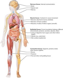
Principal Types of Body Tissue
Epithelial Tissue
Epithelial tissue forms boundaries between different environments, protects, absorbs, filters, secretes, and senses. It covers body surfaces, lines cavities, and forms glands.
Polarity: Has an apical (top) and basal (bottom) surface.
Specialized contacts: Cells are tightly joined by junctions (tight junctions, desmosomes).
Supported by connective tissue: The basal surface is attached to a basement membrane.
Avascular but innervated: Contains no blood vessels but is supplied by nerves.
Regeneration: High capacity for renewal.
Classification of Epithelial Tissue
Epithelial tissues are classified by the number of layers and cell shape:
Simple: One layer (for absorption, secretion, filtration).
Stratified: Multiple layers (for protection).
Shapes: Squamous (flat), Cuboidal (cube-shaped), Columnar (tall).
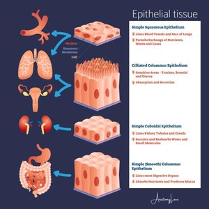
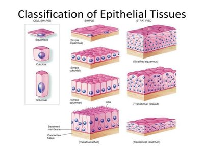
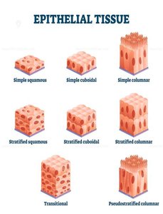
Examples of Epithelial Tissue Types
Type | Structure | Location | Function |
|---|---|---|---|
Simple Squamous | Single flat layer | Lungs, kidneys, blood vessels | Diffusion, filtration |
Simple Cuboidal | Single cube-shaped layer | Kidney tubules, glands | Secretion, absorption |
Simple Columnar | Single tall layer | Digestive tract, uterine tubes | Absorption, secretion |
Pseudostratified Columnar | Appears layered, but is not | Respiratory tract | Secretion, movement of mucus |
Stratified Squamous | Multiple flat layers | Skin, mouth, esophagus | Protection |
Stratified Cuboidal | Two layers, cube-shaped | Sweat, mammary glands | Protection |
Stratified Columnar | Multiple tall layers | Pharynx, male urethra | Protection, secretion |
Transitional | Shape-shifting | Bladder, ureters | Stretching |
Glandular Epithelia
Glands are specialized epithelial structures that secrete substances. They are classified as endocrine (ductless, secrete hormones into blood) or exocrine (secrete products into ducts or onto surfaces).
Endocrine glands: Release hormones directly into the bloodstream (e.g., thyroid, pituitary).
Exocrine glands: Release products onto body surfaces or into body cavities (e.g., sweat, salivary glands).
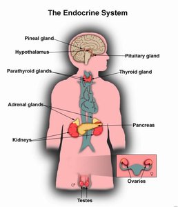
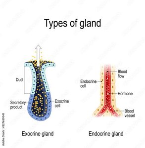
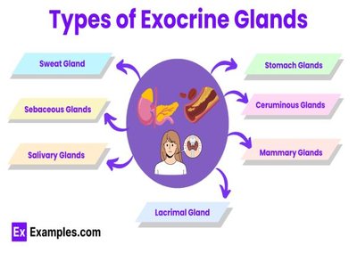
Modes of Secretion (Exocrine Glands)
Type | Mechanism | Example |
|---|---|---|
Merocrine | Secrete by exocytosis | Salivary, sweat glands |
Holocrine | Cell ruptures to release product | Sebaceous (oil) glands |
Apocrine | Apex of cell pinches off | Mammary glands (debated) |
Connective Tissue
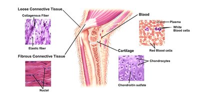
Connective tissue is the most abundant and widely distributed tissue type. It supports, binds, protects, insulates, stores energy, and transports substances throughout the body.
Major classes: Connective tissue proper, cartilage, bone, blood.
Functions: Binding/support, protection, insulation, energy storage, transport.
Muscle Tissue
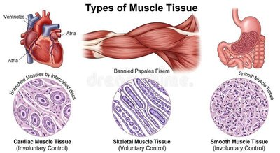
Muscle tissue is specialized for contraction and movement. It is highly vascularized and responsible for most types of body movement.
Skeletal muscle: Attached to bones, voluntary movement.
Cardiac muscle: Found in the heart, involuntary control.
Smooth muscle: Found in walls of hollow organs, involuntary control.
Nervous Tissue
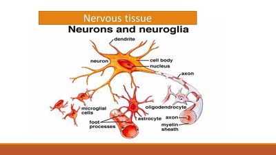
Nervous tissue is the main component of the nervous system (brain, spinal cord, nerves). It regulates and controls body functions through electrical impulses.
Neurons: Specialized cells that generate and conduct nerve impulses.
Neuroglia: Supporting cells that protect, insulate, and support neurons.
Body Membranes
Major Categories of Body Membranes
Body membranes are composed of epithelial tissue bound to underlying connective tissue. They cover surfaces, line cavities, and form protective sheets around organs.
Cutaneous membrane: The skin; dry, keratinized stratified squamous epithelium.
Mucous membranes: Line body cavities open to the exterior (digestive, respiratory, urinary tracts); moist.
Serous membranes: Line closed ventral body cavities; produce serous fluid; have parietal and visceral layers (pleura, pericardium, peritoneum).
Tissue Repair and Healing
Process of Tissue Repair
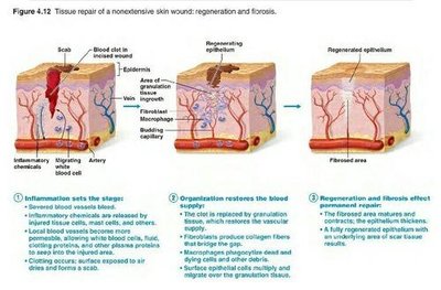
When tissues are injured, the body initiates a repair process involving inflammation, organization, and regeneration or fibrosis. The goal is to restore tissue integrity and function.
Inflammation: Inflammatory chemicals are released, blood vessels dilate, and clotting occurs to prevent further injury and infection.
Organization: The blood clot is replaced by granulation tissue, new capillaries form, fibroblasts produce collagen, and debris is removed.
Regeneration and Fibrosis: Epithelium regenerates, the scab detaches, and underlying scar tissue forms. Function is restored if regeneration predominates; otherwise, fibrosis leads to scar formation.
Regenerative Capacity of Tissues
Regenerates Well | Moderate | Poor |
|---|---|---|
Epithelial tissue Bone Areolar CT Dense irregular CT Blood-forming tissue | Smooth muscle Dense regular CT | Cardiac muscle Nervous tissue (brain/spinal cord) |
Additional info: New research is exploring ways to enhance regeneration in tissues with poor natural healing capacity, such as cardiac and nervous tissue.
