 Indietro
Chapter 4: Tissue—The Living Fabric (Anatomy & Physiology Study Notes)
Guida di studio - Note intelligenti
 Appunti personalizzati basati sui tuoi materiali, ampliati con definizioni chiave, esempi e contesto.
Tissue: The Living Fabric
Introduction to Tissues
Tissues are groups of cells with similar structure and function, forming the foundation of organs and systems in the human body. Understanding tissue types is essential for monitoring health and diagnosing conditions such as bedsores and cancer.
Histology: The study of tissues.
Four basic tissue types: Epithelial, connective, muscle, and nervous tissue.
Homeostasis: Maintained by specialized functions of individual body cells.
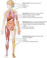
Overview of Four Basic Tissue Types
Epithelial, Connective, Muscle, and Nervous Tissues
Each tissue type has unique functions and structural characteristics:
Epithelial Tissue: Covers surfaces, lines cavities, forms glands; functions include protection, absorption, filtration, excretion, secretion, and sensory reception.
Connective Tissue: Supports, protects, binds other tissues; includes bone, cartilage, blood, and proper connective tissue.
Muscle Tissue: Responsible for movement; includes skeletal, cardiac, and smooth muscle.
Nervous Tissue: Regulates and controls body functions; consists of neurons and supporting glial cells.
Preparation of Tissue Samples for Microscopy
Fixation, Sectioning, and Staining
To study tissues under a microscope, samples must be prepared through several steps:
Fixation: Preserves tissue structure using solvents.
Sectioning: Slices tissue thinly for light or electron transmission.
Staining: Enhances contrast; light microscopy uses colored dyes, electron microscopy uses heavy metal salts.
Types of Electron Microscopy: Transmission (TEM) shows internal sections; Scanning (SEM) shows surface details.
Epithelial Tissue
Definition and Functions
Epithelial tissue forms protective sheets covering surfaces and lining cavities. It is essential for various physiological processes.
Covering and lining epithelium: Skin, linings of urogenital, digestive, and respiratory systems.
Glandular epithelium: Forms glands such as salivary glands.
Main functions: Protection, absorption, filtration, excretion, secretion, sensory reception.
Special Characteristics of Epithelial Tissues
Epithelial tissues possess five key characteristics:
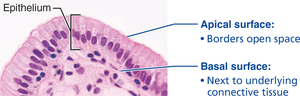
Polarity: Distinct apical (top) and basal (bottom) surfaces.
Specialized contacts: Tight junctions and desmosomes bind cells together.
Supported by connective tissue: Basement membrane (basal and reticular lamina) reinforces and defines boundaries.
Avascular but innervated: No blood vessels; nourished by diffusion; supplied by nerve fibers.
Regeneration: High capacity for renewal, especially in areas exposed to friction or hostile environments.
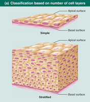
Classification of Epithelial Tissue
Epithelial tissues are classified by cell layers and cell shape:
Number of cell layers: Simple (one layer) or stratified (multiple layers).
Cell shape: Squamous (flat), cuboidal (cube-shaped), columnar (tall).
In stratified epithelia, cell shape is named according to the apical layer.
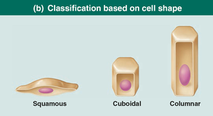
Types of Epithelial Tissue
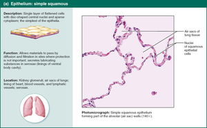
Simple Squamous Epithelium
Single layer of flattened cells; functions in rapid diffusion and filtration. Found in kidneys, lungs, blood vessels (endothelium), and serous membranes (mesothelium).
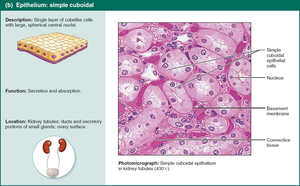
Simple Cuboidal Epithelium
Single layer of cube-shaped cells; functions in secretion and absorption. Found in kidney tubules and gland ducts.
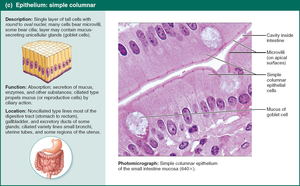
Simple Columnar Epithelium
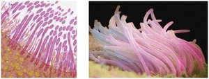
Single layer of tall cells; may have microvilli or cilia. Functions in absorption and secretion of mucus, enzymes, and other substances. Found in digestive tract, gallbladder, bronchi, and uterine tubes.
Pseudostratified Columnar Epithelium
Appears multi-layered but is actually a single layer; often ciliated. Functions in secretion and movement of mucus. Found in upper respiratory tract and ducts of large glands.
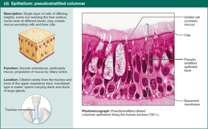
Summary Table: Epithelial Tissue Types
Type | Structure | Function | Location |
|---|---|---|---|
Simple Squamous | Single layer, flat | Diffusion, filtration | Kidney, lungs, vessels |
Simple Cuboidal | Single layer, cube | Secretion, absorption | Kidney tubules, glands |
Simple Columnar | Single layer, tall | Absorption, secretion | Digestive tract, bronchi |
Pseudostratified Columnar | Single layer, varied height | Mucus secretion, movement | Respiratory tract |
Stratified Squamous | Multiple layers, flat apical | Protection | Skin, mouth, esophagus |
Transitional | Multiple layers, shape changes | Stretching | Bladder, ureters |
Glandular Epithelia
Types of Glands
Endocrine glands: Ductless, secrete hormones into interstitial fluid and blood.
Exocrine glands: Secrete products onto surfaces or into cavities via ducts (e.g., sweat, oil, salivary glands).
Unicellular glands: Goblet cells produce mucin.
Multicellular glands: Composed of ducts and secretory units; classified by structure (simple/compound, tubular/alveolar) and mode of secretion (merocrine, holocrine, apocrine).
Connective Tissue
Definition and Functions
Connective tissue is the most abundant tissue type, providing support, protection, insulation, storage, and transport.
Four main classes: Connective tissue proper, cartilage, bone, blood.
Extracellular matrix: Nonliving material suspending cells; allows tissue to bear weight and tension.
Common origin: All arise from mesenchyme (embryonic tissue).
Structural Elements of Connective Tissue
Ground substance: Gel-like material with interstitial fluid, cell adhesion proteins, and proteoglycans.
Fibers: Collagen (strength), elastic (stretch/recoil), reticular (support).
Cells: Immature "-blast" cells (secrete matrix), mature "-cyte" cells (maintain matrix), adipocytes (fat storage), leukocytes (immune response), mast cells (inflammation), macrophages (phagocytosis).
Muscle Tissue
Types and Functions
Muscle tissue is specialized for contraction and movement. It is highly vascularized and contains myofilaments (actin and myosin).
Skeletal muscle: Voluntary, striated, multiple nuclei, moves bones.
Cardiac muscle: Involuntary, striated, one nucleus, found in heart, intercalated discs.
Smooth muscle: Involuntary, non-striated, spindle-shaped, one nucleus, found in walls of hollow organs.
Nervous Tissue
Structure and Function
Nervous tissue forms the brain, spinal cord, and nerves, regulating and controlling body functions.
Neurons: Respond to stimuli (dendrites), transmit electrical signals (axons).
Glial cells (neuroglia): Support, insulate, and protect neurons.
Membranes
Types of Membranes
Membranes are organs composed of more than one tissue type, covering and lining body surfaces.
Cutaneous membrane: Skin; keratinized stratified squamous epithelium attached to connective tissue; dry membrane.
Mucous membrane: Lines cavities open to exterior; moist, may secrete mucus; found in digestive, respiratory, urogenital tracts.
Serous membrane: Lines closed ventral body cavities; simple squamous epithelium (mesothelium) on areolar connective tissue; moist, secretes serous fluid.
Tissue Repair
Steps in Tissue Repair
Tissue repair is activated by inflammatory and immune responses when barriers are compromised. Repair occurs in three steps:
Inflammation: Release of chemicals, dilation of blood vessels, increased permeability, clotting.
Organization: Blood clot replaced by granulation tissue, regeneration of epithelium, collagen fibers bridge gap, debris removed.
Regeneration and fibrosis: Scab detaches, fibrous tissue matures, epithelium thickens, scar tissue may remain.
Regenerative Capacity of Tissues
High regeneration: Epithelial, bone, areolar, dense irregular, blood-forming tissue.
Moderate regeneration: Smooth muscle, dense regular connective tissue.
Low/no regeneration: Cardiac muscle, nervous tissue (brain, spinal cord).
Developmental Aspects of Tissues
Primary Germ Layers
Embryonic development forms three primary germ layers:
Ectoderm: Forms nerve tissue.
Mesoderm: Forms muscle and connective tissues.
Endoderm: Contributes to epithelial tissues.
Tissues function well through youth and middle age, but aging leads to thinning epithelia, less efficient repair, atrophy, and increased cancer risk.
