 Indietro
Chapter 4: Tissue—The Living Fabric (Anatomy & Physiology Study Notes)
Guida di studio - Note intelligenti
 Appunti personalizzati basati sui tuoi materiali, ampliati con definizioni chiave, esempi e contesto.
Chapter 4: Tissue—The Living Fabric
Section 1: Introduction to Tissues
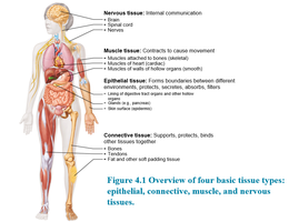
Tissues are groups of cells with similar structure and function, forming the "living fabric" of the body. The four basic tissue types are epithelial, connective, muscle, and nervous tissues, each with distinct roles in the human body.
Epithelial Tissue: Forms boundaries between different environments, protects, secretes, absorbs, and filters.
Connective Tissue: Supports, protects, and binds other tissues together.
Muscle Tissue: Contracts to cause movement.
Nervous Tissue: Enables internal communication.
Section 2: Epithelial Tissue
Epithelial tissue covers body surfaces and lines cavities, forming protective barriers and facilitating absorption, secretion, and filtration. It is characterized by five distinguishing features: polarity, specialized contacts, support by connective tissue, avascularity, and regeneration.
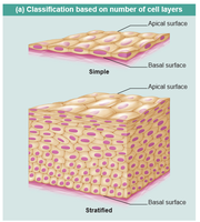
Polarity: Epithelial cells have an apical surface (exposed to body exterior or cavity) and a basal surface (attached to underlying connective tissue).
Specialized Contacts: Cells are tightly joined by junctions to form continuous sheets.
Support by Connective Tissue: The basal surface is anchored to a basement membrane.
Avascular but Innervated: Epithelial tissues lack blood vessels but are supplied by nerves.
Regeneration: High capacity for renewal, especially after injury.
Classification of Epithelia
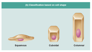
Epithelia are classified based on the number of cell layers and cell shape:
Number of Layers:
Simple Epithelium: Single layer of cells; functions in absorption, secretion, and filtration.
Stratified Epithelium: Multiple layers; provides protection.
Cell Shape:
Squamous: Flat, scale-like cells.
Cuboidal: Cube-shaped cells.
Columnar: Tall, column-like cells.
Examples of Epithelial Types
Each epithelial type has specific functions and locations:
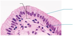
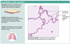
Simple Squamous Epithelium: Allows diffusion and filtration; found in air sacs of lungs, lining of heart, blood vessels.
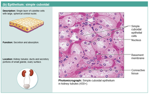
Simple Cuboidal Epithelium: Secretion and absorption; found in kidney tubules, ducts, and secretory portions of small glands.
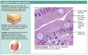
Simple Columnar Epithelium: Absorption, secretion of mucus and enzymes; lines most of the digestive tract, gallbladder, and some ducts.
Section 3: Glandular Epithelium
Glandular epithelia form glands that secrete substances. Glands are classified as endocrine (secrete hormones into blood) or exocrine (secrete products onto body surfaces or into cavities).
Goblet Cells: Unicellular glands that secrete mucus.
Modes of Secretion:
Merocrine: Secrete by exocytosis (e.g., sweat glands).
Holocrine: Entire cell ruptures, releasing secretions and cell fragments (e.g., sebaceous glands).
Section 4: Connective Tissue
Connective tissue is the most abundant and widely distributed tissue in the body. It supports, protects, and binds other tissues. All connective tissues share three basic components: fibers, ground substance, and cells.
Fibers: Collagen (strength), elastic (flexibility), reticular (support).
Ground Substance: Fills space between cells and fibers; composed of interstitial fluid, proteins, and polysaccharides.
Cells: Fibroblasts, chondrocytes, osteocytes, and others depending on tissue type.
Comparison of Classes of Connective Tissues
Type | Main Function | Location |
|---|---|---|
Connective Tissue Proper | Binding, support, storage | Fat, tendons, ligaments |
Cartilage | Support, flexibility | Joints, ear, nose |
Bone | Support, protection | Skeletal system |
Blood | Transport | Blood vessels |
Structural Elements of Connective Tissue
Extracellular Matrix: Nonliving material that separates cells; provides structural support.
Fibers: Collagen, elastic, and reticular fibers.
Cells: Fibroblasts (produce fibers), adipocytes (store fat), immune cells (defense).
Section 5: Types of Connective Tissue
Connective tissue is classified into connective tissue proper, cartilage, bone, and blood. Each has unique structural and functional properties.
Loose Connective Tissue: Areolar, adipose, and reticular tissues; provide support and cushioning.
Dense Connective Tissue: Regular, irregular, and elastic tissues; provide strength and flexibility.
Cartilage: Hyaline, elastic, and fibrocartilage; support and flexibility in joints and structures.
Bone: Hard, calcified matrix; supports and protects.
Blood: Fluid tissue; transports nutrients, gases, and wastes.
Section 6: Muscle Tissue
Muscle tissue is specialized for contraction and movement. There are three types: skeletal, cardiac, and smooth muscle.
Skeletal Muscle: Voluntary movement; attached to bones.
Cardiac Muscle: Involuntary; found in the heart.
Smooth Muscle: Involuntary; found in walls of hollow organs.
Section 7: Nervous Tissue
Nervous tissue is responsible for internal communication. It consists of neurons (transmit electrical signals) and supporting cells (glia).
Neurons: Specialized cells for receiving and transmitting signals.
Glial Cells: Support, protect, and nourish neurons.
Section 8: Body Membranes
Body membranes cover surfaces, line cavities, and form protective sheets. The main types are mucous, serous, and cutaneous membranes.
Mucous Membranes: Line body cavities open to the exterior; secrete mucus.
Serous Membranes: Line closed cavities; secrete serous fluid.
Cutaneous Membrane: The skin; protects the body.
Section 9: Tissue Repair and Healing
Tissue repair involves restoring tissue integrity after injury. The process includes inflammation, organization, and regeneration/fibrosis.
Step 1: Inflammation sets the stage by releasing chemicals, making blood vessels leaky, and forming a clot and scab.
Step 2: Organization restores blood supply as granulation tissue forms, fibroblasts produce collagen, and macrophages clear debris.
Step 3: Regeneration and Fibrosis effect permanent repair as epithelium regenerates and scar tissue forms.
Regenerative Capacity of Different Tissues
High Regeneration: Epithelial tissues, bone, areolar, dense irregular connective tissue, blood-forming tissues.
Moderate Regeneration: Smooth muscle, dense regular connective tissue.
Low/No Regeneration: Cardiac muscle, nervous tissue in brain and spinal cord.
Clinical—Homeostatic Imbalance
Cartilage: Avascular cartilage heals slowly; may calcify or ossify with age.
Scar Tissue: Excessive scarring can impair organ function, block substances, and cause progressive failure, especially in the heart.
Chapter 4 Key Takeaways
Tissues are fundamental units of structure and function in the body.
Epithelial tissue forms protective barriers and glands.
Connective tissue supports, binds, and protects.
Muscle tissue enables movement.
Nervous tissue facilitates communication.
Tissue repair involves inflammation, organization, and regeneration/fibrosis.
