 Indietro
Chapter 5: The Integumentary System – Anatomy & Physiology Study Notes
Guida di studio - Note intelligenti
 Appunti personalizzati basati sui tuoi materiali, ampliati con definizioni chiave, esempi e contesto.
The Integumentary System
Overview
The integumentary system is composed of the skin and its derivatives, including hair, nails, sweat glands, and sebaceous (oil) glands. It serves as the body's primary barrier against the external environment and performs several essential functions.
Protection: Shields internal tissues from mechanical, chemical, and biological threats.
Body temperature regulation: Maintains homeostasis through sweat production and blood flow.
Cutaneous sensations: Detects touch, pressure, pain, and temperature.
Metabolic functions: Synthesizes vitamin D and other metabolic processes.
Blood reservoir: Stores blood in the skin's vessels.
Excretion of wastes: Removes waste products via sweat.
Structure of the Skin
Skin Regions
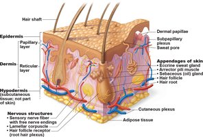
The skin consists of two main regions and an associated layer:
Epidermis: The superficial, avascular layer made of epithelial tissue.
Dermis: The deeper, vascular layer composed of fibrous connective tissue.
Hypodermis (superficial fascia): A subcutaneous layer mainly of adipose tissue, not technically part of the skin but shares some functions.
Epidermis
Cell Types in the Epidermis
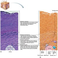
The epidermis is primarily made of keratinized stratified squamous epithelium and contains four main cell types:
Keratinocytes: Produce keratin, the protein that provides protective properties. These cells are tightly connected by desmosomes and are constantly shed.
Melanocytes: Located in the deepest epidermis, produce melanin pigment to protect against UV damage.
Dendritic (Langerhans) cells: Immune cells that patrol the epidermis.
Tactile (Merkel) cells: Sensory receptors for touch.
Layers of the Epidermis
The epidermis is organized into distinct layers (strata):
Stratum basale: Deepest layer, single row of stem cells, site of active mitosis.
Stratum spinosum: Several layers thick, contains prekeratin filaments and dendritic cells.
Stratum granulosum: Four to six layers, cells flatten and keratinization begins.
Stratum lucidum: Only in thick skin (hands, feet), thin band of dead keratinocytes.
Stratum corneum: 20–30 rows of dead, keratinized cells, provides protection.
Dermis
Structure and Function
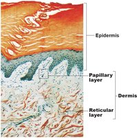
The dermis is a strong, flexible connective tissue layer containing fibroblasts, macrophages, mast cells, and white blood cells. It houses nerves, blood vessels, lymphatic vessels, hair follicles, oil glands, and sweat glands.
Dermal Layers
Papillary layer: Superficial, areolar connective tissue with dermal papillae that contain capillary loops, nerve endings, and touch receptors.
Reticular layer: Deep, dense irregular connective tissue, provides strength, elasticity, and hydration.
Clinical Note: Stretch Marks and Blisters
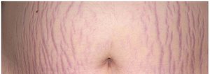
Striae (stretch marks): Result from extreme stretching, causing dermal tears.
Blisters: Fluid-filled pockets separating epidermal and dermal layers due to acute trauma.
Skin Color
Pigments Contributing to Skin Color
Melanin: Produced by melanocytes, shields DNA from UV, responsible for brown to black hues.
Carotene: Yellow to orange pigment, accumulates in stratum corneum and hypodermis, can be converted to vitamin A.
Hemoglobin: Gives pinkish hue to fair skin due to transparency and low melanin.
Clinical Note: Skin Color Changes
Cyanosis: Blue skin, indicates low oxygenation.
Erythema: Redness, due to fever, inflammation, allergy.
Pallor: Pale color, associated with anemia, low blood pressure, fear.
Jaundice: Yellow cast, indicates liver disorders.
Hair
Structure and Function
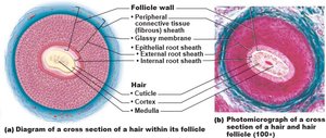
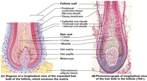
Hair consists of dead, keratinized cells and is absent from palms, soles, lips, nipples, and parts of external genitalia. Functions include protection, sensory input, and regulation of heat loss.
Regions and Parts of Hair
Shaft: Visible part above scalp, keratinization complete.
Root: Embedded in scalp, keratinization ongoing.
Hair Shaft Structure
Medulla: Central core with large cells and air spaces.
Cortex: Layers of flattened cells surrounding medulla.
Cuticle: Outermost layer of overlapping single cells.
Skin Appendages: Sweat and Oil Glands
Sweat Glands
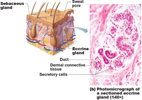
Eccrine (merocrine) sweat glands: Most numerous, abundant on palms, soles, and forehead, function in thermoregulation.
Apocrine sweat glands: Confined to axillary and anogenital areas, secrete viscous sweat, begin functioning at puberty.
Modified apocrine glands: Ceruminous glands (earwax), mammary glands (milk).
Sebaceous (Oil) Glands
Distribution: Found everywhere except palms and soles.
Function: Secrete sebum into hair follicles, bactericidal, softens hair and skin.
Activity: Stimulated by hormones, especially androgens, active after puberty.
Skin Disorders: Cancer and Burns
Skin Cancer
Basal cell carcinoma: Most common, least malignant, arises from stratum basale, cured by excision.
Squamous cell carcinoma: Second most common, can metastasize, arises from stratum spinosum, treated by surgery or radiation.
Melanoma: Most dangerous, cancer of melanocytes, highly metastatic, resistant to chemotherapy. Early detection is critical (ABCD rule: Asymmetry, Border, Color, Diameter).
Burns
First-degree: Epidermal damage only, redness, swelling, pain.
Second-degree: Epidermal and upper dermal damage, blisters.
Third-degree: Entire skin thickness destroyed, area may appear white, red, or blackened, no pain due to nerve destruction, requires skin grafting.
Immediate threat: Dehydration and electrolyte imbalance, leading to renal shutdown and circulatory shock.
Summary Table: Layers of the Skin
Layer | Location | Main Features |
|---|---|---|
Stratum basale | Deepest epidermal layer | Single row of stem cells, active mitosis |
Stratum spinosum | Above basale | Several cell layers, prekeratin filaments, dendritic cells |
Stratum granulosum | Above spinosum | Keratinization begins, lamellar granules |
Stratum lucidum | Only in thick skin | Thin, clear, dead keratinocytes |
Stratum corneum | Superficial | 20–30 rows of dead, keratinized cells |
Summary Table: Types of Skin Cancer
Type | Origin | Features | Treatment |
|---|---|---|---|
Basal cell carcinoma | Stratum basale | Least malignant, most common | Surgical excision |
Squamous cell carcinoma | Stratum spinosum | Can metastasize, scaly papule | Surgery, radiation |
Melanoma | Melanocytes | Most dangerous, highly metastatic | Surgery, immunotherapy |
Summary Table: Types of Burns
Degree | Layers Affected | Symptoms |
|---|---|---|
First-degree | Epidermis | Redness, pain, swelling |
Second-degree | Epidermis & upper dermis | Blisters, pain |
Third-degree | Entire skin | No pain, color changes, skin grafting needed |
Additional info: The notes expand on brief points with academic context, definitions, and examples to ensure completeness and clarity for exam preparation.
