 Indietro
Chapter 7: The Nervous System – Mini-Textbook Study Guide
Guida di studio - Note intelligenti
 Appunti personalizzati basati sui tuoi materiali, ampliati con definizioni chiave, esempi e contesto.
Nervous System Overview
Main Functions and Core Sequence
The nervous system is the body's primary controlling and communicating system, operating through rapid electrical impulses. Its three main functions are:
Sensory input: Gathers information by monitoring changes (stimuli) inside and outside the body.
Integration: Processes and interprets sensory input, deciding what action is needed.
Motor output: Responds to integrated stimuli by activating muscles or glands (effectors).
The sequence is: Sensory input → Integration → Motor output.
Structural and Functional Classification
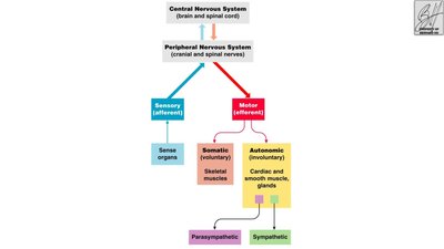
Central Nervous System (CNS): Command center consisting of the brain and spinal cord.
Peripheral Nervous System (PNS): Nerves extending from the CNS; includes cranial and spinal nerves.
Sensory (afferent) division: Carries information to the CNS.
Motor (efferent) division: Carries impulses away from the CNS to effectors.
Somatic nervous system: Voluntary control of skeletal muscles.
Autonomic nervous system: Involuntary control of smooth/cardiac muscle and glands; subdivided into sympathetic and parasympathetic divisions.
Nervous Tissue: Support Cells (Neuroglia)
Types and Functions of Neuroglia
Neuroglia (glial cells) are support cells in the CNS and PNS. They do not transmit electrical impulses and retain the ability to divide.
Support Cell | Location | Function |
|---|---|---|
Astrocytes | CNS | Anchor neurons to capillaries; regulate material exchange. |
Microglia | CNS | Dispose of debris. |
Ependymal cells | CNS | Line cavities; circulate cerebrospinal fluid (CSF). |
Oligodendrocytes | CNS | Produce myelin sheaths. |
Satellite cells | PNS | Protect neuron cell bodies. |
Schwann cells | PNS | Form myelin sheaths. |
Oligodendrocytes = CNS myelin; Schwann cells = PNS myelin.
Neuron Anatomy and Terminology
Structure and Function of Neurons
Neurons are specialized cells for transmitting electrical signals. Their major regions are:
Cell body: Contains nucleus; metabolic center.
Dendrites: Conduct impulses toward the cell body; numerous per neuron.
Axon: Conducts impulses away from the cell body; one per neuron.
Axonal terminals: Branches at axon end; contain neurotransmitter vesicles.
Synaptic cleft: Gap between adjacent neurons.
Synapse: Junction between nerves.
Myelin sheath is a fatty covering that insulates axons and increases impulse transmission speed. Nodes of Ranvier are gaps in the myelin sheath.
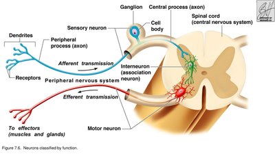
CNS terminology: Cell bodies = nuclei; nerve fibers = tracts. PNS terminology: Cell bodies = ganglia; nerve fibers = nerves.
Gray matter: Unmyelinated fibers. White matter: Myelinated fibers (tracts); cell bodies (nuclei).
Classification of Neurons
Functional Classification
Sensory (afferent) neurons: Carry impulses from sensory receptors to CNS; include cutaneous sense organs and proprioceptors.
Motor (efferent) neurons: Carry impulses from CNS to muscles/glands.
Interneurons (association neurons): Connect sensory and motor neurons within CNS.
Proprioceptors detect stretch/tension in muscles, tendons, and joints.
Structural Classification
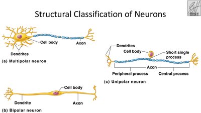
Multipolar neuron: Many dendrites, one axon.
Bipolar neuron: One dendrite, one axon.
Unipolar neuron: Single short process with peripheral and central branches.
Reflexes and Reflex Arcs
Types and Elements of Reflexes
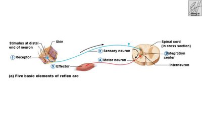
Reflexes are rapid, predictable, involuntary responses to stimuli, occurring over neural pathways called reflex arcs. Types include:
Somatic reflex: Stimulates skeletal muscles (e.g., withdrawal from heat).
Autonomic reflex: Regulates smooth muscle, heart, glands, blood pressure, digestion.
The five elements of a reflex arc are:
Sensory receptor – reacts to stimulus
Sensory neuron – carries message to CNS
Integration center – processes information
Motor neuron – carries message to effector
Effector organ – muscle or gland stimulated
Central Nervous System: Brain
Major Brain Regions
Cerebral hemispheres (cerebrum): Paired superior parts; >50% of brain mass.
Diencephalon
Brain stem
Cerebellum
Gyri: Elevated ridges. Sulci: Shallow grooves. Fissures: Deep grooves dividing lobes.
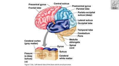
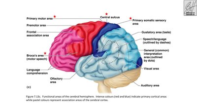
Cerebral Cortex Functional Areas
Primary somatic sensory area: Receives impulses from sensory receptors (pain, cold, touch).
Primary motor area: Sends impulses to skeletal muscles.
Broca's area: Speech/vocalization.
Gustatory area: Taste.
Visual area: Vision.
Auditory area: Hearing.
Olfactory area: Smell.
Interpretation areas: Speech/language and comprehension.
Cerebral White Matter
Fiber tracts: Deep to gray matter; transmit impulses to/from cortex.
Corpus callosum: Connects hemispheres; transmits impulses between them.
Diencephalon, Brain Stem, and Cerebellum
Diencephalon
Thalamus: Sensory relay station; transfers impulses for localization/interpretation.
Hypothalamus: Autonomic center; regulates temperature, metabolism, drives; part of limbic system; pituitary gland attached.
Epithalamus: Pineal body; choroid plexus forms CSF.
Brain Stem
Midbrain: Mostly tracts.
Pons: Fiber tracts; nuclei for breathing control.
Medulla oblongata: Control centers for heart rate, blood pressure, breathing, swallowing, vomiting.
Cerebellum
Two hemispheres; convoluted surfaces.
Outer cortex = gray matter; inner region = white matter.
Involuntary coordination for balance/equilibrium.
Interprets equilibrium info from ears, eyes, proprioceptors.
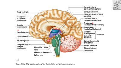
Protection of the Central Nervous System
Meninges
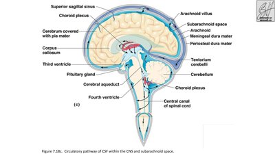
The meninges are connective tissue membranes surrounding the CNS:
Dura mater: Outermost, double-layered covering.
Arachnoid mater: Middle, web-like layer; villi protrude through dura.
Pia mater: Internal layer; clings to brain/spinal cord surface.
Order: Dura mater → Arachnoid mater → Pia mater (D-A-P).
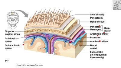
Cerebrospinal Fluid (CSF)
Similar to blood plasma.
Formed by choroid plexuses.
Watery cushion protecting brain from trauma.
Circulates in subarachnoid space and central canal; reabsorbed at subdural sinuses.
Blood-Brain Barrier
Allows passage of water, glucose, essential amino acids, fats, respiratory gases, alcohol, nicotine, anesthesia.
Excludes urea, toxins, proteins, most drugs.
Spinal Cord
Anatomy and Function
Extends from foramen magnum to L1-L2 vertebra.
Two-way conduction pathway to/from brain.
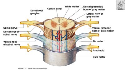
Internal gray matter: Cell bodies; dorsal (posterior) and anterior (ventral) horns; surrounds central canal.
Central canal: Filled with CSF.
Exterior white matter: Conduction tracts; divided into dorsal, lateral, ventral columns.
Meninges: Cover spinal cord.
Spinal nerves: Leave at each vertebra level.
Dorsal root: Associated with dorsal root ganglia (cell bodies outside CNS).
Ventral root: Contains axons.
Quick-Review Table: Nervous System Terms
Term | Must-Know Idea |
|---|---|
Sensory input | Gathers information about stimuli |
Integration | Processes/interprets sensory input |
Motor output | Activates muscles or glands |
CNS | Brain + spinal cord |
PNS | Cranial + spinal nerves outside CNS |
Afferent | To CNS |
Efferent | Away from CNS |
Somatic motor | Voluntary skeletal muscle control |
Autonomic | Involuntary smooth/cardiac muscle + glands |
Astrocyte | Anchors neurons; regulates exchange |
Microglia | Debris disposal |
Ependymal | Line CNS cavities; circulate CSF |
Oligodendrocyte | CNS myelin |
Schwann cell | PNS myelin |
Satellite cell | Protects neuron cell bodies in PNS |
Dendrite | Impulse toward cell body |
Axon | Impulse away from cell body |
Node of Ranvier | Gap in myelin sheath |
CNS cell-body cluster | Nucleus |
PNS cell-body cluster | Ganglion |
CNS nerve-fiber bundle | Tract |
PNS nerve-fiber bundle | Nerve |
Sensory neuron | Receptor → CNS |
Motor neuron | CNS → muscle/gland/viscera |
Interneuron | Connects sensory and motor neurons in CNS |
Reflex arc | Receptor → sensory → integration → motor → effector |
Thalamus | Sensory relay station |
Hypothalamus | Autonomic center; temperature/metabolism/drives |
Medulla oblongata | Heart rate, BP, breathing, swallowing, vomiting |
Cerebellum | Balance/equilibrium + coordination |
Broca’s area | Speech/vocalization |
Corpus callosum | Connects cerebral hemispheres |
Meninges | Dura → arachnoid → pia |
Choroid plexus | Forms CSF |
CSF | Watery cushion protecting CNS |
Practice Questions (with Answers)
Which part of the nervous system contains the brain and spinal cord? Answer: CNS
Which division carries nerve impulses toward the CNS? Answer: Afferent
Which glial cell forms myelin in the PNS? Answer: Schwann cell
Which glial cell disposes of debris? Answer: Microglia
Dendrites conduct impulses in which direction? Answer: Toward the cell body
A bundle of nerve fibers in the CNS is called a: Answer: Tract
Which neuron connects sensory and motor neurons in the CNS? Answer: Interneuron
Which is the first element of a reflex arc? Answer: Sensory receptor
Which reflex type stimulates skeletal muscles? Answer: Somatic reflex
Which structure is the relay station for sensory impulses? Answer: Thalamus
Which area sends impulses to skeletal muscles? Answer: Primary motor area
Broca's area is associated with: Answer: Speaking/vocalizing
Which structure connects the two cerebral hemispheres? Answer: Corpus callosum
Which brain-stem region contains control centers for heart rate and blood pressure? Answer: Medulla oblongata
Which structure provides involuntary coordination for balance and equilibrium? Answer: Cerebellum
Put the meninges in order from outside to inside: Answer: Dura → arachnoid → pia
What forms cerebrospinal fluid according to the slides? Answer: Choroid plexuses
The spinal cord extends to approximately which vertebral level? Answer: L1-L2
In the spinal cord, gray matter is mainly: Answer: Internal
The dorsal root is associated with the: Answer: Dorsal root ganglion
Neuroglia transmit electrical impulses. Answer: False
Nodes of Ranvier are gaps in the myelin sheath. Answer: True
The somatic nervous system allows voluntary control of skeletal muscles. Answer: True
The pia mater is the outermost meningeal layer. Answer: False
The central canal of the spinal cord contains cerebrospinal fluid. Answer: True
Additional info: This guide covers the core concepts and terminology for Chapter 7: The Nervous System, suitable for exam preparation in Anatomy & Physiology college courses.
