 Indietro
Chapter 8: Articulations – Structured Study Notes for Anatomy & Physiology
Guida di studio - Note intelligenti
 Appunti personalizzati basati sui tuoi materiali, ampliati con definizioni chiave, esempi e contesto.
Articulations (Joints)
Functions of Joints
Joints, also known as articulations, are the locations where two bones meet. They serve several essential functions in the human body:
Enable Movement: Muscles and tendons exert force across joints, allowing for a wide range of movements.
Provide Stability: Joints with limited or no mobility are highly stable, protecting underlying structures (e.g., skull joints).
Allow Long Bones to Lengthen: The epiphyseal plate is a temporary joint that enables bone growth during development.
Classification of Joints
Functional Classification
Joints are classified by the degree of movement they allow:
Synarthrosis: No movement; most stable (e.g., skull sutures).
Amphiarthrosis: Small amount of movement; significant stability (e.g., intervertebral discs).
Diarthrosis: Freely moveable; least stable (e.g., shoulder joint).
Structural Classification
Structural classification is based on the connective tissue linking bones and the presence of a joint cavity:
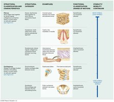
Fibrous Joints: Dense regular collagenous connective tissue; no joint space; functionally synarthroses or amphiarthroses.
Cartilaginous Joints: Cartilage between bones; no joint space; functionally synarthroses or amphiarthroses.
Synovial Joints: Joint cavity filled with fluid; functionally diarthroses.
Fibrous Joints
Types of Fibrous Joints
Fibrous joints are stabilized by collagen fibers and permit little movement:
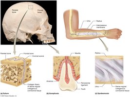
Sutures: Found between skull bones; interlocking projections held by short collagen fibers; very stable synarthroses. Fusion forms synostosis.
Gomphoses: Joint between a tooth and its alveolus; stabilized by the periodontal ligament; synarthroses.
Syndesmoses: Bones joined by an interosseous membrane or ligament; found between radius/ulna and tibia/fibula; amphiarthroses.
Cartilaginous Joints
Types of Cartilaginous Joints
Cartilaginous joints lack a cavity and allow limited movement:
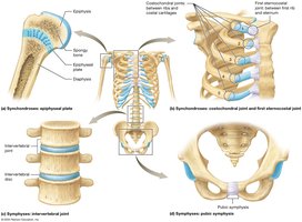
Synchondroses: Bones united by hyaline cartilage; synarthroses. Examples: epiphyseal plate, first sternocostal joint, costochondral joints.
Symphyses: Bones united by fibrocartilage pad; amphiarthroses. Examples: intervertebral joints/discs, pubic symphysis.
Epiphyseal Plate Fractures
The epiphyseal plate is a weak area in a child's skeleton. Fractures can cause limb length discrepancies, deformities, and early arthritis. Treatment ranges from immobilization to surgery.
Synovial Joints
Structure of Synovial Joints
Synovial joints are characterized by a joint cavity and are the most mobile type:
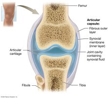
Articular Capsule: Double-layered; outer fibrous layer (dense irregular connective tissue) and inner synovial membrane (loose connective tissue).
Synovial Fluid: Lubricates, supplies nutrients, removes wastes, and absorbs shock.
Articular Cartilage: Hyaline cartilage covers bone surfaces; reduces friction and absorbs shock; avascular.
Other Components: Adipose tissue (padding), blood vessels, and nerves (pain and proprioception).
Stabilizing and Supportive Structures
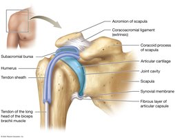
Ligaments: Connect bone to bone; intrinsic (within capsule) or extrinsic (outside capsule).
Tendons: Connect muscle to bone; stabilize joints via muscle tone.
Bursae: Synovial fluid-filled sacs that reduce friction in high-stress areas.
Tendon Sheaths: Elongated bursae surrounding tendons in high-stress regions.
Bursitis
Bursitis is inflammation of a bursa, often due to trauma, repetitive movement, or arthritis. Symptoms include pain, swelling, and warmth. Treatment includes rest, ice, compression, medication, and sometimes steroid injections.
Arthritis
Osteoarthritis: Wear and tear, injuries, aging.
Rheumatoid Arthritis: Autoimmune destruction of joints.
Gouty Arthritis: Inflammatory reaction to uric acid crystals.
Functional Classes of Synovial Joints
Axes of Motion
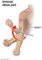
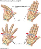
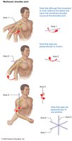
Synovial joints are classified by the number of axes around which bones move:
Nonaxial: Motion in one or more planes, not around an axis.
Uniaxial: Motion around one axis (e.g., elbow).
Biaxial: Motion around two axes (e.g., metacarpophalangeal joints).
Multiaxial (Triaxial): Motion around three axes (e.g., shoulder).
Movements at Synovial Joints
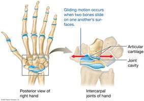
Gliding Movements
Sliding motion between bone surfaces; nonaxial.
Angular Movements
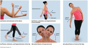
Flexion: Decreases angle between bones.
Extension: Increases angle between bones.
Hyperextension: Extension beyond anatomical position.
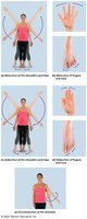
Abduction: Moves body part away from midline.
Adduction: Moves body part toward midline.
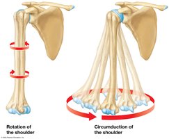
Circumduction: Distal bone moves in a cone-shaped motion.
Rotational Movements
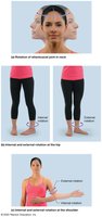
Internal (Medial) Rotation: Rotates toward midline.
External (Lateral) Rotation: Rotates away from midline.
Special Movements
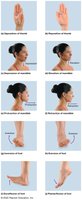
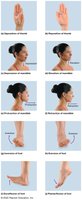
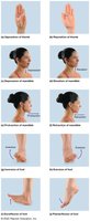
Opposition/Reposition: Thumb movement across palm and return.
Depression/Elevation: Inferior and superior movement.
Protraction/Retraction: Anterior and posterior movement.
Inversion/Eversion: Medial and lateral rotation of foot.
Dorsiflexion/Plantarflexion: Decrease/increase angle between foot and tibia.
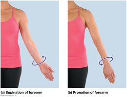
Supination/Pronation: Forearm rotation; palm faces anteriorly or posteriorly.
Range of Motion
Range of motion is the amount of movement a joint can perform. Nonaxial joints have the smallest range, multiaxial joints the greatest.
Structural Classes of Synovial Joints
Types of Synovial Joints
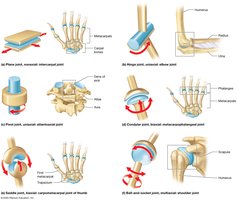
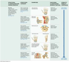
Plane Joint: Nonaxial; flat surfaces.
Hinge Joint: Uniaxial; convex fits into concave.
Pivot Joint: Uniaxial; rounded fits into groove.
Condylar (Ellipsoid) Joint: Biaxial; oval convex fits into shallow concave.
Saddle Joint: Biaxial; each surface has convex and concave regions.
Ball-and-Socket Joint: Multiaxial; spherical fits into cup/socket.
Joint Classification and Stability vs. Mobility
Fibrous and Cartilaginous Joints
Synovial Joints
Specific Hinge Joints: Elbow and Knee
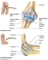
The Elbow Joint
Humeroulnar Joint: Trochlea of humerus and trochlear notch of ulna.
Humeroradial Joint: Capitulum of humerus and head of radius.
Ligaments: Radial (lateral) collateral, ulnar (medial) collateral, anular ligament.
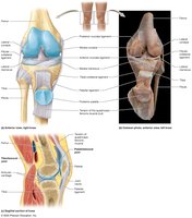
The Knee Joint
Tibiofemoral Joint: Femoral and tibial condyles.
Patellofemoral Joint: Patella and patellar surface of femur.
Menisci: Medial and lateral fibrocartilage pads.
Ligaments: Tibial (medial) collateral, fibular (lateral) collateral, anterior cruciate (ACL), posterior cruciate (PCL).
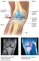
Knee Injuries and the Unhappy Triad
Lateral blows to the knee can rupture the tibial collateral ligament, lateral meniscus, and ACL, known as the "Unhappy Triad." Surgery and physical therapy are required for recovery.


Specific Ball-and-Socket Joints: Shoulder and Hip
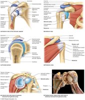
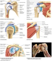
The Shoulder (Glenohumeral Joint)
Articulating Surfaces: Humeral head and glenoid cavity.
Capsule Reinforcement: Biceps brachii tendon, coracohumeral ligament, glenohumeral ligaments, subacromial and subscapular bursae.
Rotator Cuff: Subscapularis, supraspinatus, infraspinatus, teres minor.
Glenoid Labrum: Fibrocartilaginous ring for stability.
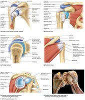
Shoulder Dislocations
Dislocation involves displacement of the humeral head from the glenoid cavity. Most occur through the inferior part of the anterior capsule. Common in falls and contact sports.

The Hip (Coxal Joint)
Articulating Surfaces: Acetabulum and femoral head.
Acetabular Labrum: Fibrocartilage ring for stability.
Capsule and Muscles: Strong capsule and powerful muscles.
Ligaments: Iliofemoral, ischiofemoral, pubofemoral, ligament of the head of femur.
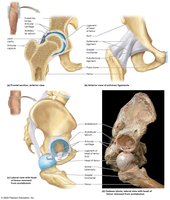
Hip Joint Replacement Surgery
Hip replacement replaces damaged joint with a prosthetic device. Total replacement reconstructs both femoral head and acetabulum; partial replaces only femoral head. Recovery includes physical therapy.


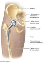
Summary Table: Joint Classification
Fibrous and Cartilaginous Joints
Structural Category | Example | Functional Classification | Stability vs. Mobility |
|---|---|---|---|
Suture | Skull | Synarthrosis | Most stable |
Gomphosis | Tooth in alveolus | Synarthrosis | Stable |
Syndesmosis | Radius/ulna, tibia/fibula | Amphiarthrosis | Moderate stability |
Synchondrosis | Epiphyseal plate, sternocostal joint | Synarthrosis | Stable |
Symphysis | Intervertebral disc, pubic symphysis | Amphiarthrosis | Moderate stability |
Synovial Joints
Structural Category | Example | Functional Classification | Stability vs. Mobility |
|---|---|---|---|
Plane | Intercarpal joints | Nonaxial | Least mobility |
Hinge | Elbow, knee | Uniaxial | Moderate mobility |
Pivot | Atlantoaxial joint | Uniaxial | Moderate mobility |
Condylar | Metacarpophalangeal joints | Biaxial | Greater mobility |
Saddle | Thumb joint | Biaxial | Greater mobility |
Ball-and-Socket | Shoulder, hip | Multiaxial | Greatest mobility |
Key Terms and Concepts
Articulation: Joint between two bones.
Synarthrosis: Immovable joint.
Amphiarthrosis: Slightly movable joint.
Diarthrosis: Freely movable joint.
Synovial Fluid: Lubricating fluid in synovial joints.
Ligament: Connects bone to bone.
Tendon: Connects muscle to bone.
Bursa: Fluid-filled sac reducing friction.
Meniscus: Fibrocartilage pad in knee.
Labrum: Fibrocartilage ring in shoulder and hip.
Formulas and Equations
While joint movement is not typically described by equations, the following formula is relevant for range of motion:
Range of Motion (ROM):
Examples and Applications
Example: The shoulder joint allows circumduction, enabling the arm to move in a circular pattern.
Application: Hip replacement surgery restores mobility in patients with severe arthritis or trauma.
Additional info: Academic context was added to clarify joint classifications, movement types, and clinical relevance.
