 Indietro
Chapter 8: Joints – Classification, Structure, and Function
Guida di studio - Note intelligenti
 Appunti personalizzati basati sui tuoi materiali, ampliati con definizioni chiave, esempi e contesto.
Joints: Classification and Function
Definition and Overview
Joints, also known as articulations, are sites where two or more bones meet. They are essential for movement and provide structural support to the skeleton. Joints are classified based on their structural composition and functional capacity.
Structural Classification: Based on what holds the joint together and whether a cavity is present.
Functional Classification: Based on the degree of movement allowed at the joint.
Structural Types:
Fibrous Joints
Cartilaginous Joints
Synovial Joints
Functional Types:
Synarthroses: Immovable joints
Amphiarthroses: Slightly movable joints
Diarthroses: Freely movable joints
Fibrous Joints
Structure and Types
Fibrous joints are united by fibrous connective tissue and lack a joint cavity, resulting in minimal to no movement.
Sutures: Found only between bones of the skull; immovable.
Syndesmoses: Bones connected by ligaments or interosseous membranes; allow limited movement.
Gomphoses: Peg-in-socket joints, such as the connection between a tooth and its bony socket.
Cartilaginous Joints
Structure and Types
Cartilaginous joints are united by cartilage and also lack a joint cavity.
Synchondroses: Bones joined by hyaline cartilage; examples include epiphyseal plates and joints between the first seven ribs and the sternum.
Symphyses: Articular surfaces covered with hyaline cartilage and a plate of fibrocartilage; provide strength and flexibility (e.g., pubic symphysis, intervertebral joints).
Synovial Joints
General Structure and Characteristics
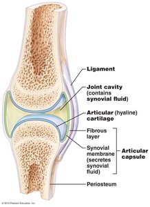
Synovial joints are the most common and freely movable joints in the body. They possess a joint cavity filled with synovial fluid and are characterized by six key features:
Articular Cartilage: Hyaline cartilage covers opposing bone surfaces, providing cushioning.
Joint Cavity: A fluid-filled space (synovial cavity) between articulating bones.
Articular Capsule: Double-layered capsule with an outer fibrous layer (dense irregular CT) and an inner synovial membrane (loose CT).
Synovial Fluid: Egg-white consistency fluid that reduces friction and nourishes cartilage.
Reinforcing Ligaments: Strengthen and maintain bone alignment.
Nerves & Blood Vessels: Nerves monitor joint position and stretch; blood vessels supply nutrients.
Bursae and Tendon Sheaths
Bursae and tendon sheaths are structures that reduce friction in synovial joints.
Bursa: Sac lined with synovial membrane, found where ligaments, muscles, or tendons rub against bone.
Tendon Sheath: Elongated bursa that wraps around a tendon.
Factors Influencing Synovial Joint Stability
Synovial joints allow significant movement but are less stable than fibrous or cartilaginous joints. Stability is influenced by:
Articular Surfaces: Deep ball-and-socket joints are more stable.
Ligaments: More ligaments provide greater strength; overstretched ligaments remain stretched.
Muscle Tone: Taut tendons crossing joints are crucial for stability, especially in the shoulder, knee, and foot arches.
Common Joint Injuries
Types of Injuries
Cartilage Injuries: Common in the knee; slow repair due to poor blood supply; may require arthroscopic surgery.
Dislocations: Bones forced out of normal position; repeat dislocations are common due to stretched ligaments.
Sprains: Partial ligament tears heal slowly; complete tears require surgical intervention.
Movements Allowed by Synovial Joints
Axes of Movement
Synovial joints allow movements along one (monoaxial), two (biaxial), or three (multiaxial) axes, corresponding to the coronal, sagittal, and transverse planes.
Types of Synovial Joints and Their Movements
The shape of articulating surfaces determines the types of movements permitted:
Plane Joint: Flat surfaces allow gliding movements (e.g., intercarpal joints).
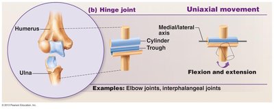
Hinge Joint: Cylinder fits into a trough, allowing flexion and extension (e.g., elbow joint).
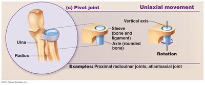
Pivot Joint: Rounded bone fits into a ring, allowing rotation (e.g., proximal radioulnar joint, atlantoaxial joint).
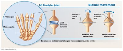
Condylar Joint: Oval articular surfaces allow movement in two planes (e.g., metacarpophalangeal joints).
Saddle Joint: Saddle-shaped surfaces permit greater freedom of movement (e.g., thumb joint).
Ball-and-Socket Joint: Spherical head fits into a cup-like socket, allowing multiaxial movement (e.g., shoulder and hip joints).
Special Movements
Examples of Special Movements
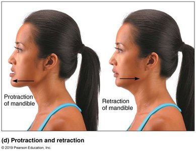
Protraction and Retraction: Moving a body part forward or backward (e.g., mandible).
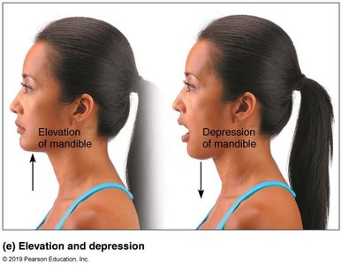
Elevation and Depression: Raising or lowering a body part (e.g., mandible).
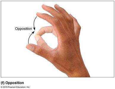
Opposition: Movement of the thumb to touch the fingertips.
Major Synovial Joints
Knee Joint
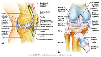
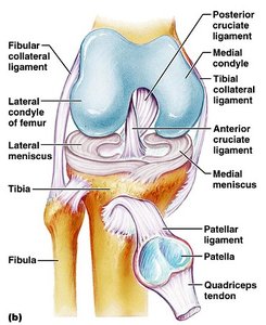
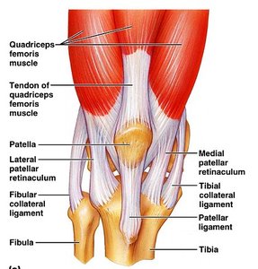
The knee is a complex joint allowing flexion, extension, and slight rotation. It consists of three joints: femoropatellar, lateral tibiofemoral, and medial tibiofemoral. The joint is stabilized by ligaments, menisci, and bursae.
Ligaments: Patellar, collateral, and cruciate ligaments.
Menisci: Medial and lateral fibrocartilage pads.
Bursae: Prepatellar, suprapatellar, and others reduce friction.
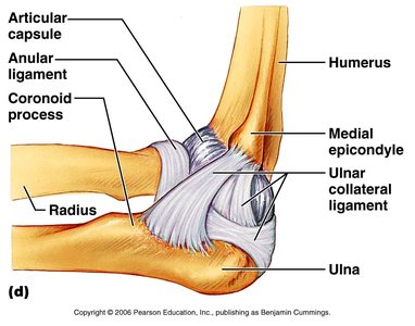
Elbow Joint
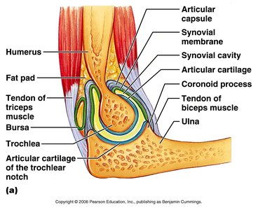
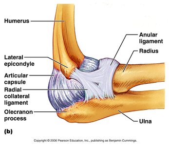
The elbow is a hinge joint primarily formed by the articulation of the humerus and ulna. It is stabilized by collateral ligaments and the annular ligament, which allows rotation of the radius during pronation and supination.
Articular Capsule: Encloses the joint.
Collateral Ligaments: Radial and ulnar collateral ligaments provide stability.
Annular Ligament: Encircles the head of the radius.
Shoulder Joint (Glenohumeral Joint)
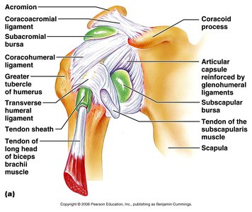
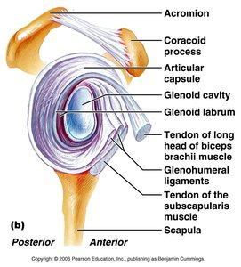
The shoulder is a ball-and-socket joint with a shallow socket (glenoid cavity) broadened by the glenoid labrum. Stability is provided mainly by the tendons of the rotator cuff muscles.
Ligaments: Coracohumeral and glenohumeral ligaments.
Bursae: Subacromial and subscapular bursae.
Rotator Cuff: Muscles and tendons reinforce the joint.
Hip Joint
The hip is a deep ball-and-socket joint with a strong capsule and several important ligaments. The acetabulum is the socket, and the head of the femur is the ball.
Ligamentum Teres: Intracapsular ligament; its function in humans is unclear.
Extracapsular Ligaments: Iliofemoral, pubofemoral, and ischiofemoral ligaments.
Temporomandibular Joint (TMJ)
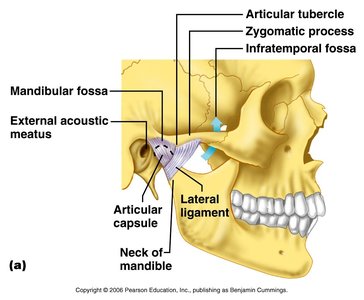
The TMJ is a modified hinge joint formed by the articulation of the mandibular condyle with the mandibular fossa and articular tubercle of the temporal bone. It is stabilized by a lateral ligament and contains an articular disk dividing the synovial cavity.
Movements: Initial jaw opening is a hinge movement, followed by sliding of the articular disk.
Summary Table: Classification of Joints
Structural Type | Functional Type | Examples | Movement |
|---|---|---|---|
Fibrous | Synarthroses | Sutures (skull) | Immovable |
Fibrous | Amphiarthroses | Syndesmoses (radius/ulna) | Limited movement |
Fibrous | Synarthroses | Gomphoses (teeth) | Immovable |
Cartilaginous | Synarthroses | Synchondroses (epiphyseal plate) | Immovable |
Cartilaginous | Amphiarthroses | Symphyses (pubic symphysis) | Slightly movable |
Synovial | Diarthroses | Shoulder, knee, hip | Freely movable |
Key Terms and Definitions
Articular Cartilage: Hyaline cartilage covering bone surfaces in synovial joints.
Synovial Fluid: Lubricating fluid within the joint cavity.
Bursa: Fluid-filled sac reducing friction.
Ligament: Connective tissue connecting bones.
Meniscus: Fibrocartilage pad in the knee.
Additional info: Academic context was added to clarify joint types, structure, and function, and to provide definitions and examples for exam preparation.
