 Indietro
Chapter 9: Joints – Structure, Function, and Clinical Relevance
Guida di studio - Note intelligenti
 Appunti personalizzati basati sui tuoi materiali, ampliati con definizioni chiave, esempi e contesto.
Joints (Articulations)
Introduction to Joints
Joints, also known as articulations, are the locations where two bones meet and where body movement occurs. The structure of a joint determines its function, and there is a tradeoff between strength and mobility: the more mobile a joint, the less stable it tends to be.
Classification of Joints
Structural and Functional Classification
Joints are classified by both their anatomical structure and their functional range of motion:
Structural Classification: Fibrous, Cartilaginous, Bony, Synovial
Functional Classification: Synarthrosis (immovable), Amphiarthrosis (slightly movable), Diarthrosis (freely movable)
Synarthrosis (Immovable Joints)
Synarthroses are very strong joints where the edges of bones may touch or interlock. They can be fibrous or cartilaginous and include:
Suture: Found only between bones of the skull, bound by dense fibrous connective tissue.
Gomphosis: Binds teeth to bony sockets via the periodontal ligament.
Synchondrosis: Rigid cartilaginous bridge, e.g., between vertebrosternal ribs and sternum.
Synostosis: Created when two bones fuse, e.g., metopic suture of frontal bone.
Amphiarthrosis (Slightly Movable Joints)
Amphiarthroses are more movable than synarthroses but stronger than diarthroses. They may be fibrous or cartilaginous:
Syndesmosis: Bones connected by a ligament.
Symphysis: Bones connected by fibrocartilage.
Diarthrosis (Freely Movable Joints)
Diarthroses are synovial joints, which are freely movable and found at the ends of long bones. They are surrounded by a joint capsule and contain synovial fluid.
Synovial Joints
Basic Structure of Synovial Joints
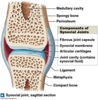
Synovial joints are characterized by their mobility and are stabilized by accessory structures. The main components include:
Joint capsule (articular capsule): Encloses the joint cavity.
Synovial membrane: Produces synovial fluid.
Synovial fluid: Lubricates, distributes nutrients, and absorbs shock.
Articular cartilage: Covers surfaces, prevents bone contact.
Accessory Structures
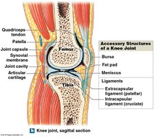
Cartilages (Meniscus): Fibrocartilage pad between bones.
Fat pads: Adipose tissue covered by synovial membrane, protects articular cartilages.
Ligaments: Support and strengthen joints; sprains occur when collagen fibers are torn.
Tendons: Attach muscles around the joint.
Bursae: Small pockets of synovial fluid that cushion areas where tendons or ligaments rub against other tissues.
Movements at Synovial Joints
Planes and Types of Movement
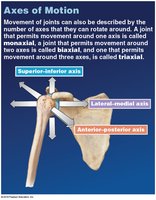
Movements are described by the plane or direction and the relationship between structures. Joints may be:
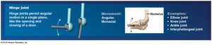
Monaxial: Movement in one plane (e.g., elbow)
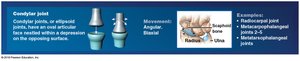
Biaxial: Movement in two planes (e.g., wrist)
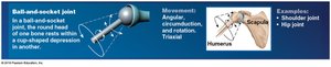
Triaxial: Movement in three planes (e.g., shoulder)
Types of Movement
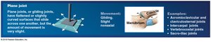
Gliding: Two flat surfaces slide past each other (e.g., carpal bones).
Angular: Includes flexion, extension, hyperextension, abduction, adduction, and circumduction.
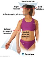
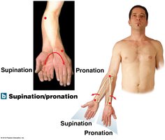
Rotational: Includes medial and lateral rotation, pronation, and supination.
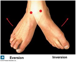
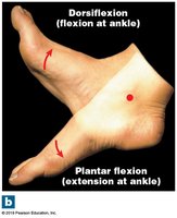
Special Movements: Inversion, eversion, dorsiflexion, plantar flexion, opposition, reposition, protraction, retraction, depression, elevation, lateral flexion.
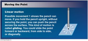
Gliding Movement
Occurs when two flat surfaces slide past each other.
Angular Movement
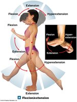
Flexion decreases the angle between bones; extension increases it; hyperextension extends past anatomical position.
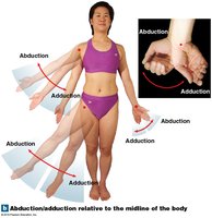
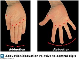
Abduction moves a limb away from the midline; adduction moves it toward the midline.
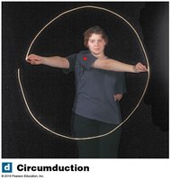
Circumduction
A circular movement without rotation.
Rotational Movement
Rotation occurs around the longitudinal axis. Pronation rotates the forearm so the palm faces posteriorly; supination turns the palm anteriorly.
Special Movements
Inversion/Eversion: Twisting the sole of the foot medially/laterally.
Dorsiflexion/Plantar Flexion: Flexion/extension at the ankle.
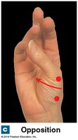
Opposition/Reposition: Movement of thumb toward/away from fingers.
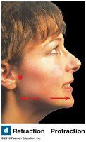
Protraction/Retraction: Moving a structure forward/backward.
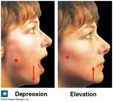
Depression/Elevation: Moving a structure down/up.
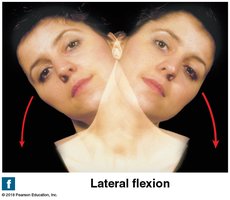
Lateral Flexion: Bending vertebral column to the side.
Classification of Synovial Joints
Synovial joints are classified by their shape and movement:
Type | Movement | Example |
|---|---|---|
Plane (gliding) | Nonaxial, limited | Intercarpal joints |
Hinge | Monaxial, angular | Elbow, knee |
Condylar (ellipsoid) | Biaxial, angular | Radiocarpal joint |
Saddle | Biaxial, angular | First carpometacarpal joint |
Pivot | Monaxial, rotation | Atlanto-axial joint |
Ball-and-socket | Triaxial, angular/circumduction/rotation | Shoulder, hip |


Intervertebral Joints
Structure and Function
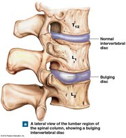
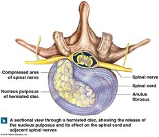
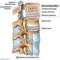
Intervertebral joints include synovial joints between articular processes and symphyses between vertebral bodies. Intervertebral discs separate vertebral bodies and consist of:
Anulus fibrosus: Tough outer layer of fibrocartilage.
Nucleus pulposus: Elastic, gelatinous core that absorbs shocks.
Vertebral end plates: Cartilage covering disc surfaces.
Intervertebral Ligaments
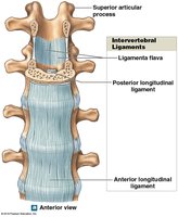
Ligamenta flava: Connect laminae of adjacent vertebrae.
Posterior longitudinal ligament: Connects posterior surfaces of vertebral bodies.
Anterior longitudinal ligament: Connects anterior surfaces of vertebral bodies.
Interspinous ligaments: Connect spinous processes.
Supraspinous ligament: Connects tips of spinous processes, continuous with ligamentum nuchae.
Elbow and Knee Joints
Elbow Joint
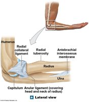
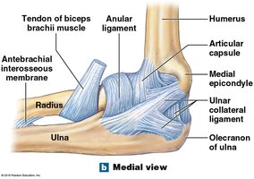
The elbow is a hinge joint involving the humerus, radius, and ulna. The humero-ulnar joint is the largest and strongest, while the humeroradial joint is smaller. The elbow is stabilized by several ligaments:
Radial collateral ligament
Anular ligament
Ulnar collateral ligament
Knee Joint
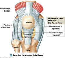
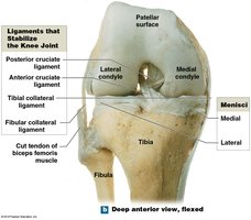
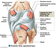
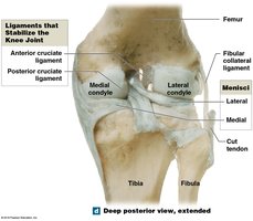
The knee is a complex hinge joint transferring weight from femur to tibia. It has three articulations and is stabilized by menisci and seven major ligaments:
Patellar ligament
Two popliteal ligaments
Anterior and posterior cruciate ligaments
Tibial collateral ligament
Fibular collateral ligament
Shoulder and Hip Joints
Shoulder Joint (Glenohumeral Joint)
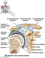
The shoulder is a ball-and-socket diarthrosis with the greatest range of motion. It is stabilized by muscles, tendons, ligaments, and the glenoid labrum.
Acromioclavicular, coracoclavicular, coraco-acromial, coracohumeral, glenohumeral ligaments
Rotator cuff muscles: Supraspinatus, infraspinatus, teres minor, subscapularis
Bursae: Subdeltoid, subcoracoid, subacromial, subscapular
Hip Joint
The hip is a strong ball-and-socket diarthrosis between the head of the femur and the acetabulum. The acetabular labrum increases depth and seals in synovial fluid. Ligaments include:
Iliofemoral
Pubofemoral
Ischiofemoral
Transverse acetabular
Ligament of the femoral head
Effects of Aging on Joints
Degenerative Changes and Clinical Problems
Aging leads to degenerative changes in joints, including:
Rheumatism: Pain and stiffness in the musculoskeletal system.
Arthritis: Inflammation of joints, including osteoarthritis (wear and tear), rheumatoid arthritis (immune attack), and gouty arthritis (uric acid crystals).
Joint immobilization: Reduces synovial fluid flow, causing arthritis symptoms.
Bone mass decreases: Increased risk of fractures.
Integration with Other Systems
Functional Relationships
The skeletal system interacts with other body systems:
Bone remodeling: Involves osteoblasts (formation) and osteoclasts (recycling).
Factors: Age, physical stress, hormones, calcium/phosphorus uptake, genetics, environment.
Muscles: Attach to bones for movement.
Endocrine system: Controls bone growth and metabolism.
Digestive/urinary systems: Provide minerals for bone growth.
Skeleton: Serves as a mineral reserve.
Summary Table: Joint Types and Movements
Joint Type | Movement | Example |
|---|---|---|
Plane (gliding) | Sliding, nonaxial | Intercarpal joints |
Hinge | Angular, monaxial | Elbow, knee |
Condylar | Angular, biaxial | Radiocarpal joint |
Saddle | Angular, biaxial | First carpometacarpal joint |
Pivot | Rotation, monaxial | Atlanto-axial joint |
Ball-and-socket | Angular, circumduction, rotation, triaxial | Shoulder, hip |
Additional info: The notes have been expanded with academic context to ensure completeness and clarity for exam preparation.
