 Indietro
Chapter 9: Muscles & Muscle Tissue – Structure, Function, and Organization
Guida di studio - Note intelligenti
 Appunti personalizzati basati sui tuoi materiali, ampliati con definizioni chiave, esempi e contesto.
Overview of Muscles
Muscle Tissue and Its Importance
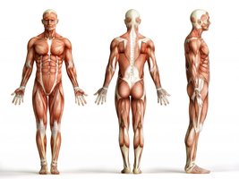
Muscles compose nearly half of the body’s mass and are essential for transforming chemical energy (ATP) into mechanical energy, enabling force generation. Muscles can only pull, not push, and are responsible for movement, posture, joint stabilization, and heat production.
Key Point: Muscles are specialized for contraction and force generation.
Key Point: Muscle tissue is classified into three types: skeletal, cardiac, and smooth.
Types of Muscle Tissue
Classification and Characteristics
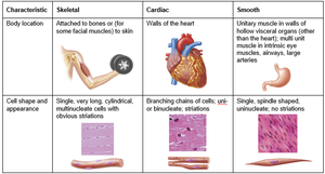
There are three types of muscle tissue, each with distinct structure and function:
Skeletal Muscle: Attached to bones or skin, voluntary, striated, long cylindrical fibers.
Cardiac Muscle: Found in the heart, involuntary, striated, branching cells.
Smooth Muscle: Located in walls of hollow organs, involuntary, non-striated, spindle-shaped cells.
Characteristic | Skeletal | Cardiac | Smooth |
|---|---|---|---|
Body location | Attached to bones or skin | Walls of the heart | Walls of hollow organs |
Cell shape & appearance | Long, cylindrical, multinucleate, striated | Branching, uni- or binucleate, striated | Spindle-shaped, uninucleate, no striations |
Skeletal Muscle Tissue
Structure and Function
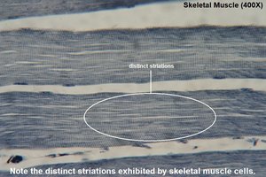
Skeletal muscle fibers are the longest muscle cells, ranging from a few millimeters to over 30 cm. They have visible striations due to the organized arrangement of proteins and are under voluntary control, except for reflexes.
Key Words: Skeletal, striated, voluntary.
Example: Muscles used for walking, lifting, and facial expressions.
Characteristics of Muscle Tissue
Four Main Properties
All muscle tissues share four fundamental characteristics:
Excitability (Responsiveness): Ability to receive and respond to stimuli, usually from the nervous system.
Contractility: Ability to shorten forcibly when stimulated, producing force.
Extensibility: Ability to be stretched or lengthened.
Elasticity: Ability to recoil to resting length after being stretched or contracted.
Functions of Skeletal Muscle
Major Roles in the Body
Skeletal muscles perform four essential functions:
Movement: Contract to produce locomotion and manipulation.
Posture: Maintain body position and stability.
Joint Stabilization: Reinforce and stabilize joints.
Heat Generation: Produce heat to maintain body temperature (e.g., shivering).
Skeletal Muscle Structure
Components of Skeletal Muscle
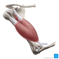
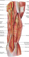
Skeletal muscle is an organ composed of muscle fibers, nerves, blood vessels, and connective tissue.
Nerve and Blood Supply: Each muscle receives a nerve, artery, and veins for control, oxygen/nutrient delivery, and waste removal.
Connective Tissue Sheaths: Layers of connective tissue organize and support muscle fibers.
Attachments: Muscles attach to bones or skin at origin and insertion points.
Connective Tissue Sheaths
Organization and Support
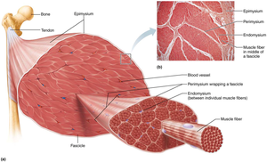
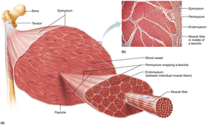
Skeletal muscle is organized by three main connective tissue sheaths:
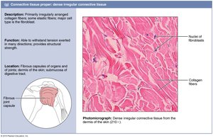
Epimysium: Outermost layer, dense irregular connective tissue, surrounds entire muscle.
Perimysium: Surrounds groups of muscle fibers (fascicles), fibrous connective tissue.
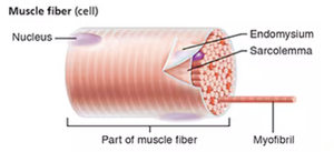
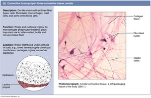
Endomysium: Fine areolar connective tissue, surrounds each individual muscle fiber.
Muscle Attachments
Origin and Insertion
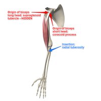
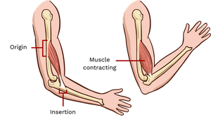
Muscles span joints and attach to bones or skin.
Origin: Attachment to immovable or less movable bone.
Insertion: Attachment to movable bone.
Direct Attachment: Epimysium fused to periosteum or perichondrium.
Indirect Attachment: Connective tissue extends as tendon or aponeurosis.
Skeletal Muscle Fiber Structure
Cellular Components
Skeletal muscle fibers are long, cylindrical cells with multiple nuclei.
Sarcolemma: Plasma membrane of muscle fiber.
Sarcoplasm: Cytoplasm containing glycosomes (glycogen storage) and myoglobin (oxygen storage).
Myofibrils: Rodlike elements responsible for contraction.
Sarcoplasmic Reticulum: Modified smooth ER, stores and releases calcium.
T Tubules: Extensions of sarcolemma, carry action potentials deep into the fiber.
Myofibrils and Sarcomeres
Functional Units of Contraction
Myofibrils are densely packed and account for most of the muscle cell volume.
Striations: Alternating light (I band) and dark (A band) regions.
Sarcomere: Smallest functional unit, region between two Z discs, contains A band and half of I band at each end.
Myofilaments: Actin (thin) and myosin (thick) filaments arranged in a precise pattern.
Molecular Composition of Myofilaments
Thick and Thin Filaments
Thick Filaments: Composed of myosin molecules with globular heads that bind to actin during contraction.
Thin Filaments: Composed of actin, tropomyosin, and troponin. Actin has binding sites for myosin; tropomyosin and troponin regulate access to these sites.
Elastic Filament: Titin protein holds thick filaments in place and provides elasticity.
Muscle Contraction Mechanism
Sequence of Events
Muscle contraction involves a series of steps:
Nervous system stimulates muscle fiber.
Calcium is released inside the cell.
Myosin binds to actin.
Filaments slide past each other, shortening the sarcomere.
Excitation-Contraction Coupling
Linking Electrical Signal to Contraction
An action potential travels along the sarcolemma and T tubules.
Sarcoplasmic reticulum releases Ca²⁺ into sarcoplasm.
Ca²⁺ binds to troponin, causing tropomyosin to move and expose binding sites on actin.
Myosin binds to actin, initiating contraction.
Sliding Filament Model
How Muscles Shorten
Myosin heads form cross bridges with actin.
Myosin pulls actin toward the center of the sarcomere.
Thin filaments slide past thick filaments; sarcomere shortens.
Actin and myosin filaments do not shorten, they slide past each other.
Cross Bridge Cycle
Steps in Muscle Contraction
Myosin binds to actin (cross bridge formation).
Power stroke: Myosin pulls actin toward center.
ATP binds to myosin, causing release from actin.
ATP is hydrolyzed, re-cocking myosin head.
Cycle repeats as long as calcium and ATP are present.
Muscle Relaxation
Ending Contraction
Nerve stimulation ends; acetylcholine is removed.
Calcium is pumped back into the sarcoplasmic reticulum.
Troponin returns to original shape; tropomyosin covers actin binding sites.
Cross bridges stop forming; muscle returns to relaxed state.
Motor Neuron Activation
Neuromuscular Junction and Action Potential
Motor neuron sends action potential to muscle.
Acetylcholine (ACh) is released at neuromuscular junction.
ACh binds to receptors on muscle fiber, generating action potential.
Action potential spreads across sarcolemma and into T tubules.
Ion Channels and Membrane Potential
Role in Muscle Activation
Chemically Gated Channels: Opened by neurotransmitters (e.g., ACh).
Voltage-Gated Channels: Opened by changes in membrane potential.
Resting Membrane Potential: Inside of cell is more negative (~ -90 mV).
Depolarization: Na⁺ enters, making inside more positive.
Repolarization: K⁺ exits, restoring negative potential.
Refractory Period: Cell cannot respond to another stimulus during repolarization.
Hyperpolarization: Membrane becomes more negative than resting value.
Na⁺/K⁺ Pump: Restores ion distribution after repolarization.
Summary Table: Muscle Organization
Hierarchy of Muscle Structure
Level | Description |
|---|---|
Muscle | Organ composed of fascicles |
Fascicle | Bundle of muscle fibers |
Muscle Fiber (Cell) | Long, multinucleate cell |
Myofibril | Rodlike contractile element |
Sarcomere | Functional unit of contraction |
Actin & Myosin Filaments | Protein filaments responsible for contraction |
Key Equations
Resting Membrane Potential
Resting membrane potential is maintained by ion gradients:
Na⁺/K⁺ pump equation:
Additional info:
Muscle contraction is regulated by calcium ions released from the sarcoplasmic reticulum.
ATP is essential for both contraction and relaxation phases.
Muscle fibers are organized into fascicles, which are grouped to form the whole muscle.
