 Indietro
Chromatin Structure and Regulation: DNA Packaging, Chromatin Remodeling, and X-Inactivation
Guida di studio - Note intelligenti
 Appunti personalizzati basati sui tuoi materiali, ampliati con definizioni chiave, esempi e contesto.
Chromatin Structure and DNA Packaging
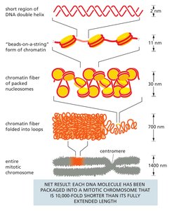
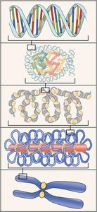
Levels of DNA Packaging
Cells must efficiently package their DNA to fit within the nucleus while maintaining accessibility for gene expression. This packaging occurs through a hierarchical series of structural levels:
DNA Double Helix: The basic structure of DNA, a 2 nm-wide double helix.
Nucleosome: DNA wraps around histone octamers, forming 'beads-on-a-string' 11 nm fibers.
Chromatin Fiber: Nucleosomes are packed into a 30 nm fiber.
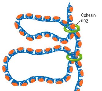
Looped Domains: Chromatin fibers fold into loops, often anchored by proteins such as cohesin.
Mitotic Chromosome: The most compact form, with DNA condensed about 10,000-fold.
Example: Each human cell contains about 2 meters of DNA, which is compacted to fit within a nucleus only a few micrometers in diameter.
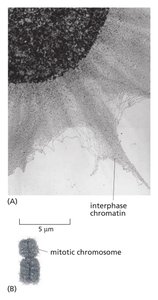
Chromatin Compaction: Interphase vs. Mitotic Chromosomes
Chromatin structure varies with the cell cycle. During interphase, chromatin is less compact, allowing gene expression and DNA replication. In mitosis, chromosomes are highly condensed for segregation.
Interphase Chromatin: Appears as a tangled mass, less condensed.
Mitotic Chromosome: Highly compact, visible as distinct structures under the microscope.
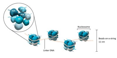
Nucleosome Structure
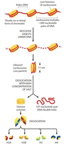
The nucleosome is the fundamental unit of chromatin, consisting of DNA wrapped around a histone octamer.
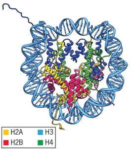
Histone Octamer: Composed of two copies each of H2A, H2B, H3, and H4.
Linker DNA: Connects adjacent nucleosomes.
Beads-on-a-string: The first level of chromatin organization.
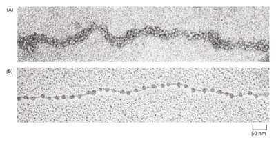
Nucleosome Disassembly
Nuclease digestion and high salt concentration can separate nucleosomes into their components:
Nuclease: Cuts linker DNA, releasing nucleosome core particles.
High Salt: Dissociates the core into histone octamers and DNA.
Histone H1: Binds linker DNA, not part of the octamer.
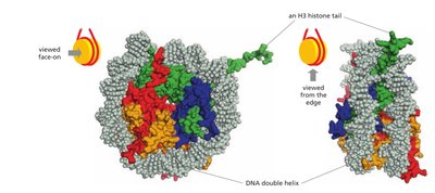
Crystal Structure of the Nucleosome
X-ray diffraction reveals the tight bending of DNA around the histone core and the projection of histone tails.
Histone Tails: Extend from the core and are sites for regulatory modifications.
DNA-Histone Interaction: The minor groove of DNA faces the histone core.
Higher-Order Chromatin Structure: Looped Domains
Chromatin fibers are further organized into looped domains, which are stabilized by structural proteins such as cohesin.
Loop Domain: Large supercoiled loops of chromatin fiber.
Cohesin: Ring-shaped protein complex that encircles the fiber at the base of each loop.
Nuclear Scaffold: Protein network anchoring loops.
Regulation of Chromatin Structure
Chromatin Remodeling
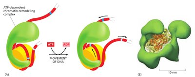
Chromatin remodeling complexes use ATP to reposition nucleosomes, altering DNA accessibility for transcription and other processes.
ATP-Dependent Remodeling: Complexes slide DNA along the histone core.
Regulatory DNA Exposure: Remodeling can expose or hide regulatory sequences.
Histone Modifications and the Histone Code
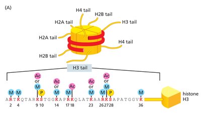
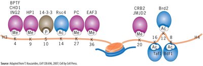
Histone tails are subject to various post-translational modifications, which regulate chromatin structure and gene activity.
Common Modifications: Acetylation, methylation, phosphorylation.
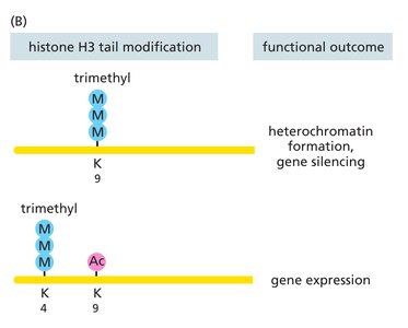
Histone Code Hypothesis: The combination of marks determines chromatin function.
Writers, Readers, Erasers: Enzymes add, recognize, or remove marks.
Euchromatin vs. Heterochromatin
Chromatin exists in two main states, affecting gene expression:
Euchromatin: Loosely packed, transcriptionally active.
Heterochromatin: Tightly packed, transcriptionally silent.
Constitutive Heterochromatin: Always condensed (e.g., centromeres, telomeres).
Facultative Heterochromatin: Condensed only in certain cells or developmental stages (e.g., inactive X chromosome).

Propagation of Heterochromatin
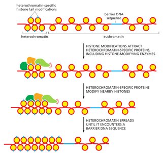
Heterochromatin spreads along the chromosome by recruiting proteins that reproduce the same histone marks on neighboring nucleosomes.
Boundary Elements: DNA sequences that prevent the spread of heterochromatin.
X-Inactivation: Epigenetic Inheritance
Mechanism of X-Inactivation
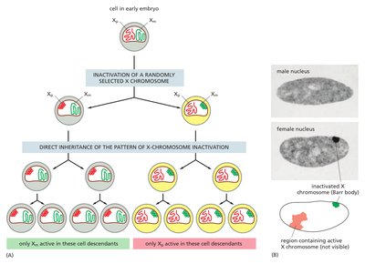
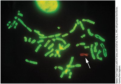
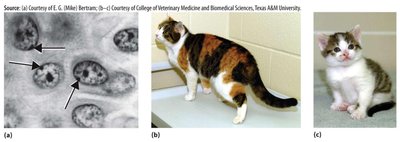
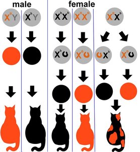
In female mammals, one X chromosome is randomly inactivated early in development, forming a Barr body. This silencing is maintained in all descendant cells, creating a mosaic pattern.
XIST RNA: Coats the chosen X chromosome and recruits silencing enzymes.
Epigenetic Inheritance: The inactive state is heritable through cell division.
Reactivation: Occurs in germ cells before meiosis.
Visible Effects: Calico Cats
Calico cats display visible mosaicism due to X-inactivation. Coat color genes on the X chromosome result in patches of different colors depending on which X is active in each cell lineage.
XX Females: Can show both black and orange patches.
XY Males: Usually show only one color.
XXY Males: Rare, can show mosaicism.
Chromatin and Disease: Rett Syndrome
Rett Syndrome and Chromatin Regulation
Rett syndrome is caused by mutations in MECP2, an X-linked gene encoding a reader of DNA methylation. The severity of the disease depends on which X chromosome is inactivated in neurons.
MECP2 Function: Binds methylated DNA, recruits histone deacetylases, compacts chromatin, and silences genes.
Epigenetic Mosaicism: Random X-inactivation leads to a mosaic of affected and unaffected neurons.
Therapeutic Approaches: Reactivating the healthy MECP2 copy or gene therapy are under investigation.
Summary and Open Questions
Chromatin Structure: DNA is packaged into nucleosomes and higher-order structures.
Regulation: Chromatin remodeling and histone modifications control gene accessibility.
Epigenetic Inheritance: Chromatin states can be inherited without changes to DNA sequence.
Disease Link: Chromatin regulation is crucial for normal development and function; its disruption can cause disease.
Additional info: The textbook 30 nm fiber is rarely seen in living cells; chromatin folding is more irregular and variable than previously thought. Cohesin is thought to extrude loops, but the exact mechanism and influence on gene expression remain active areas of research.
