 Indietro
Comprehensive Study Guide: Cardiovascular System (Anatomy & Physiology)
Guida di studio - Note intelligenti
 Appunti personalizzati basati sui tuoi materiali, ampliati con definizioni chiave, esempi e contesto.
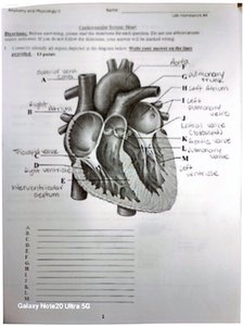
Q1. Label the anatomical structures of the heart in the diagram.
Background
Topic: Heart Anatomy
This question tests your knowledge of the internal and external structures of the heart, including chambers, valves, and major vessels. Understanding these structures is fundamental for learning about cardiac physiology and blood flow.
Key Terms:
Right/Left Atrium
Right/Left Ventricle
Aorta
Pulmonary Trunk
Superior/Inferior Vena Cava
Interventricular Septum
Valves (tricuspid, bicuspid/mitral, pulmonary, aortic)
Step-by-Step Guidance
Examine the diagram and identify the labeled points (A–L). Each label corresponds to a specific anatomical structure.
Recall the location and function of each heart chamber and vessel. For example, the right atrium receives blood from the body, while the left ventricle pumps blood to the systemic circulation.
Match the labels to the structures based on their position in the diagram. Use clues such as vessel direction and chamber wall thickness.
Review the names and functions of the heart valves, and identify their locations in the diagram.
Write the correct anatomical term for each label in the provided list.
Try solving on your own before revealing the answer!
Final Answer:
The correct anatomical terms for each label are as follows:
A: Superior vena cava
B: Right atrium
C: Right ventricle
D: Inferior vena cava
E: Left ventricle
F: Left atrium
G: Pulmonary trunk
H: Aorta
I: Pulmonary veins
J: Interventricular septum
K: Tricuspid valve
L: Bicuspid (mitral) valve
These labels correspond to the major chambers, vessels, and valves of the heart, which are essential for understanding blood flow and cardiac function.
Q2. The atria receive blood from all of the following except:
Background
Topic: Cardiac Circulation
This question tests your understanding of the sources of blood entering the atria, which is important for tracing the flow of blood through the heart.
Key Terms:
Atria
Pulmonary veins
Superior/inferior vena cava
Coronary sinus
Pulmonary artery
Step-by-Step Guidance
Recall which vessels bring blood to the right and left atria.
Identify the function of each vessel listed in the options.
Determine which vessel does not supply blood to either atrium.
Review the pathway of blood flow through the heart.
Try solving on your own before revealing the answer!
Final Answer:
The pulmonary artery does not supply blood to the atria. It carries blood away from the right ventricle to the lungs.
Q3. Which is the most likely effect of physical endurance training on a person's cardiac performance?
Background
Topic: Cardiac Physiology & Adaptation
This question examines how exercise affects heart function, particularly cardiac output and efficiency.
Key Terms:
Cardiac output
Stroke volume
Heart rate
Endurance training
Step-by-Step Guidance
Consider how regular endurance training affects the heart's ability to pump blood.
Recall the physiological adaptations that occur with exercise (e.g., increased stroke volume, decreased resting heart rate).
Evaluate the options for changes in cardiac output and heart rate.
Think about the long-term benefits of endurance training on cardiovascular health.
Try solving on your own before revealing the answer!
Final Answer:
Endurance training increases cardiac output and efficiency, often resulting in a lower resting heart rate and higher stroke volume.
Q4. The highest need for the heart due to increased workload is:
Background
Topic: Cardiac Metabolism
This question tests your understanding of what the heart requires most when it is working harder, such as during exercise.
Key Terms:
Oxygen supply
ATP production
Coronary circulation
Step-by-Step Guidance
Recall the primary energy source for cardiac muscle cells.
Consider what happens to the heart's metabolic needs during increased activity.
Identify which resource is most critical for sustaining increased workload.
Try solving on your own before revealing the answer!
Final Answer:
Oxygen is the highest need for the heart during increased workload, as it is essential for ATP production in cardiac muscle cells.
Q5. The term used to describe a backflow of blood from the left ventricle into the left atrium is:
Background
Topic: Heart Valves & Pathology
This question tests your knowledge of valve function and common cardiac pathologies related to valve insufficiency.
Key Terms:
Regurgitation
Mitral valve
Valve insufficiency
Step-by-Step Guidance
Recall the function of the mitral (bicuspid) valve between the left atrium and left ventricle.
Consider what happens if the valve does not close properly.
Identify the term used for blood flowing backward through a valve.
Try solving on your own before revealing the answer!
Final Answer:
Regurgitation is the term used to describe backflow of blood from the left ventricle into the left atrium due to mitral valve insufficiency.
Q6. Which vessel does not supply the myocardium with blood?
Background
Topic: Coronary Circulation
This question tests your understanding of the vessels responsible for supplying oxygenated blood to the heart muscle.
Key Terms:
Coronary arteries
Myocardium
Pulmonary artery
Step-by-Step Guidance
Recall which vessels are part of the coronary circulation.
Identify the function of each vessel listed in the options.
Determine which vessel does not supply blood to the myocardium.
Try solving on your own before revealing the answer!
Final Answer:
The pulmonary artery does not supply the myocardium with blood; it carries blood from the right ventricle to the lungs.
Q7. Which of the following statements about muscle tissue is true?
Background
Topic: Cardiac Muscle Physiology
This question tests your understanding of the unique properties of cardiac muscle tissue compared to other muscle types.
Key Terms:
Cardiac muscle
Intercalated discs
Automaticity
Striated muscle
Step-by-Step Guidance
Recall the structural features of cardiac muscle cells.
Identify the role of intercalated discs in cardiac muscle function.
Compare cardiac muscle to skeletal and smooth muscle.
Evaluate the statements for accuracy based on your knowledge of muscle physiology.
Try solving on your own before revealing the answer!
Final Answer:
Cardiac muscle cells are connected by intercalated discs, which allow for synchronized contraction and efficient transmission of electrical impulses.
Q8. Any abnormal cardiac rhythm is called:
Background
Topic: Cardiac Electrophysiology
This question tests your knowledge of terminology related to abnormal heart rhythms.
Key Terms:
Arrhythmia
Normal sinus rhythm
Tachycardia
Bradycardia
Step-by-Step Guidance
Recall the definition of normal sinus rhythm.
Identify the term used for any deviation from normal rhythm.
Review examples of abnormal rhythms (e.g., tachycardia, bradycardia, fibrillation).
Try solving on your own before revealing the answer!
Final Answer:
Any abnormal cardiac rhythm is called an arrhythmia.
Q9. What is indicated by a heart murmur detected between the first and second heart sounds?
Background
Topic: Heart Sounds & Valve Function
This question tests your understanding of the timing and causes of heart murmurs, which are often related to valve abnormalities.
Key Terms:
Heart murmur
Systole
Valve stenosis/regurgitation
First (S1) and second (S2) heart sounds
Step-by-Step Guidance
Recall the events that occur between S1 and S2 (ventricular systole).
Identify which valves are open and closed during this phase.
Consider what could cause a murmur during systole (e.g., stenosis or regurgitation of a semilunar valve).
Try solving on your own before revealing the answer!
Final Answer:
A murmur detected between the first and second heart sounds usually indicates stenosis or regurgitation of a semilunar valve during ventricular systole.
Q10. Which vessel carries blood from the right ventricle to the lungs?
Background
Topic: Pulmonary Circulation
This question tests your knowledge of the pathway of blood from the heart to the lungs for oxygenation.
Key Terms:
Pulmonary trunk
Pulmonary arteries
Right ventricle
Step-by-Step Guidance
Recall the direction of blood flow from the right ventricle.
Identify the vessel that carries deoxygenated blood to the lungs.
Review the anatomy of the pulmonary circulation.
Try solving on your own before revealing the answer!
Final Answer:
The pulmonary trunk carries blood from the right ventricle to the lungs, where it branches into the pulmonary arteries.
