 Indietro
Comprehensive Study Notes on Joints (Articulations) for Anatomy & Physiology
Guida di studio - Note intelligenti
 Appunti personalizzati basati sui tuoi materiali, ampliati con definizioni chiave, esempi e contesto.
Joints (Articulations)
Introduction to Joints
Joints, also known as articulations, are locations where two or more bones meet. They play a crucial role in providing movement and stability to the skeletal system. The degree of movement allowed by a joint depends on its structure and classification.
Definition: A joint is any place where adjacent bones or bone and cartilage come together to form a connection.
Function: Joints allow for movement or provide stability, depending on their type.
Relationship: Increased movement typically results in decreased stability.
Classification of Joints
Functional Classification
Joints are functionally classified based on their mobility:
Synarthrosis: Immobile joint (e.g., sutures of the skull).
Amphiarthrosis: Slightly movable joint (e.g., pubic symphysis).
Diarthrosis: Freely movable joint (e.g., shoulder joint).
Structural Classification
Structural classification is based on the material connecting the bones:
Fibrous Joints: Bones connected by fibrous connective tissue.
Cartilaginous Joints: Bones connected by cartilage.
Synovial Joints: Bones separated by a fluid-filled cavity.
Fibrous Joints
Types of Fibrous Joints
Fibrous joints are immovable and lack a joint cavity. There are three main types:
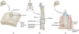
Sutures: Found between bones of the skull; dense fibrous connective tissue joins bones.
Syndesmoses: Bones connected by a band of fibrous tissue (e.g., interosseous membrane between radius and ulna).
Gomphoses: Peg-in-socket joints (e.g., teeth in their sockets).
Cartilaginous Joints
Types of Cartilaginous Joints
Cartilaginous joints are united by cartilage and lack a joint cavity. They are not highly movable.
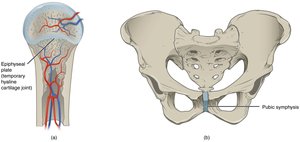
Synchondroses: Bones joined by hyaline cartilage; mostly immovable (e.g., epiphyseal plate in growing bones).
Symphyses: Bones joined by fibrocartilage; slightly movable (e.g., pubic symphysis).
Synovial Joints
General Features of Synovial Joints
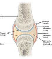
Synovial joints are the most common and freely movable joints in the body. They are characterized by the presence of a joint cavity filled with synovial fluid.
Articular Capsule: Encloses the joint cavity; consists of an outer fibrous layer and an inner synovial membrane.
Articular Cartilage: Hyaline cartilage covers the articulating surfaces, reducing friction.
Synovial Fluid: Lubricates and nourishes the joint.
Structural Features of Synovial Joints
Ligaments: Connect bones and stabilize the joint; classified as extrinsic (outside capsule) or intracapsular (inside capsule).
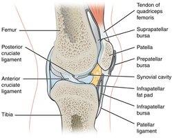
Bursae: Fluid-filled sacs that reduce friction between tissues.
Tendon Sheaths: Elongated bursae that wrap around tendons.
Types of Synovial Joints
Classification Based on Shape and Movement
Synovial joints are classified into six types based on the shape of their articulating surfaces and the movements they allow:
Pivot: Rotation around an axis (e.g., atlantoaxial joint).
Hinge: Flexion and extension (e.g., elbow).
Saddle: Allows movement in two planes (e.g., thumb).
Plane: Gliding movements (e.g., intercarpal joints).
Condylar: Flexion, extension, adduction, abduction (e.g., wrist).
Ball-and-Socket: Multiaxial movement (e.g., shoulder, hip).
Movements Allowed by Synovial Joints
Types of Movements
Synovial joints allow a wide range of movements, which are classified based on the plane and direction:
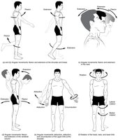
Flexion: Decreases the angle between bones.
Extension: Increases the angle between bones.
Hyperextension: Extension beyond normal range.
Abduction: Moves limb away from midline.
Adduction: Moves limb toward midline.
Circumduction: Circular movement combining flexion, extension, abduction, and adduction.
Rotation: Movement around a longitudinal axis.
Special Movements
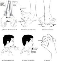
Supination and Pronation: Rotation of forearm; supination turns palm up, pronation turns palm down.
Dorsiflexion and Plantar Flexion: Movements at the ankle; dorsiflexion lifts the foot, plantar flexion points toes.
Inversion and Eversion: Inversion turns sole inward, eversion turns sole outward.
Protraction and Retraction: Anterior and posterior movement of jaw or scapula.
Elevation and Depression: Upward and downward movement of jaw or scapula.
Opposition and Reposition: Thumb movement to touch fingers and return.
Selected Synovial Joints
Shoulder (Glenohumeral) Joint
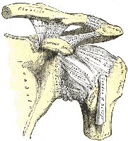
The shoulder joint is a ball-and-socket joint formed by the head of the humerus and the glenoid cavity of the scapula. It allows the greatest range of motion but is less stable.
Ligaments: Coracohumeral and glenohumeral ligaments provide support.
Knee Joint
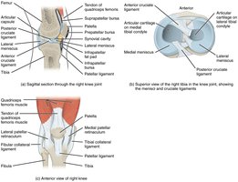
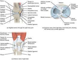
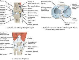
The knee is the largest joint and functions as a hinge joint. It is stabilized by several ligaments and contains menisci for shock absorption.
Extrinsic Ligaments: Fibular (lateral) and tibial (medial) collateral ligaments.
Intracapsular Ligaments: Anterior and posterior cruciate ligaments (ACL & PCL).
Menisci: Medial and lateral meniscus provide cushioning.
Knee Injuries
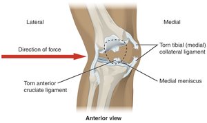
Trauma to the knee can result in the "terrible triad" injury: damage to the tibial collateral ligament, medial meniscus, and anterior cruciate ligament.
Elbow Joint
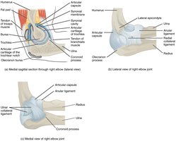
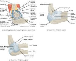
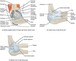
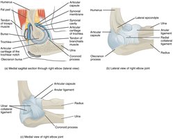
The elbow is a hinge joint formed by the humeroulnar, humeroradial, and proximal radioulnar joints. It is stabilized by ulnar and radial collateral ligaments.
Hip (Coxal) Joint
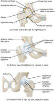
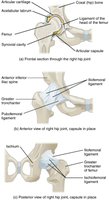
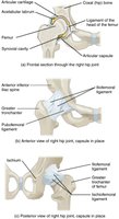
The hip is a multiaxial ball-and-socket joint between the head of the femur and the acetabulum. It is designed for strength and stability.
Ligaments: Iliofemoral, pubofemoral, and ischiofemoral ligaments spiral around the femur.
Inflammatory and Degenerative Conditions
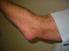
Bursitis
Bursitis is the inflammation of a bursa, causing pain, swelling, and joint stiffness. It can be acute or chronic and may result from overuse, trauma, or infection.
Tendinitis
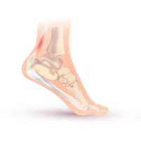
Tendinitis is the inflammation of a tendon, usually due to overuse, resulting in pain and tenderness near a joint.
Arthritis
Arthritis is a disorder involving inflammation of synovial joints. It has many forms, including osteoarthritis and rheumatoid arthritis.
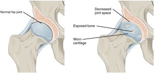
Osteoarthritis (OA)
Most common type, associated with aging and wear of articular cartilage.
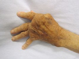
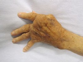
Rheumatoid Arthritis (RA)
An autoimmune disease causing inflammation of the joint capsule and synovial membrane, leading to joint deformation and disability.
Summary Table: Structural and Functional Classification of Joints
Structural Type | Functional Type | Example | Movement |
|---|---|---|---|
Fibrous (Suture) | Synarthrosis | Skull sutures | Immovable |
Fibrous (Syndesmosis) | Amphiarthrosis | Interosseous membrane | Slightly movable |
Fibrous (Gomphosis) | Synarthrosis | Tooth in socket | Immovable |
Cartilaginous (Synchondrosis) | Synarthrosis | Epiphyseal plate | Immovable |
Cartilaginous (Symphysis) | Amphiarthrosis | Pubic symphysis | Slightly movable |
Synovial (Various types) | Diarthrosis | Shoulder, hip, knee | Freely movable |
Key Equations and Concepts
Joint Stability vs. Mobility
There is an inverse relationship between joint stability and mobility:
Stability ∝ 1/Mobility
Synovial Fluid Production
Synovial fluid is produced by the synovial membrane as a plasma filtrate:
Example Applications
Jumping: Involves flexion and extension at the knee and hip joints.
Weight Lifting: Uses flexion, extension, and rotation at various synovial joints.
Swimming: Requires circumduction and rotation at shoulder and hip joints.
Additional info:
Ligaments and tendons are both composed of dense connective tissue, but ligaments connect bone to bone, while tendons connect muscle to bone.
Shoulder joint is more prone to dislocation than hip due to its greater range of motion and less structural support.
