 Indietro
Comprehensive Study Notes on Muscle Tissue and Physiology
Guida di studio - Note intelligenti
 Appunti personalizzati basati sui tuoi materiali, ampliati con definizioni chiave, esempi e contesto.
Muscle Tissue Types
Overview of Muscle Tissue
Muscle tissue is one of the four primary tissue types in the human body. It is essential for movement, posture, and various physiological functions. Muscle organs are composed of muscle tissue, nerves, blood vessels, and connective tissues.
Key Point: Muscle tissue is classified into three types: skeletal, cardiac, and smooth.
Key Point: Each type has unique structural and functional characteristics.
Example: Skeletal muscle enables voluntary movement, cardiac muscle powers the heart, and smooth muscle controls involuntary movements in internal organs.
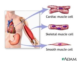
Classification of Muscle Tissue
Muscle tissue can be classified based on control, location, and structure.
Skeletal Muscle: Voluntary, striated, attached to bones, responsible for movement and posture.
Cardiac Muscle: Involuntary, striated, found in the heart, responsible for pumping blood.
Smooth Muscle: Involuntary, non-striated, found in walls of internal organs, responsible for movements like peristalsis.
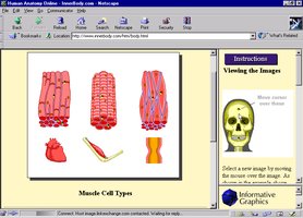
Skeletal Muscle
Structure and Function
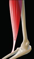
Skeletal muscle is composed of long, cylindrical, multinucleated fibers. It is responsible for voluntary movements and is attached to bones by tendons.
Key Point: Skeletal muscle fibers are striated and organized in antagonistic pairs.
Key Point: Each fiber contains myofibrils, which are the contractile elements.
Example: Muscles in the limbs facilitate locomotion and maintain posture.
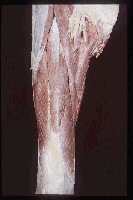
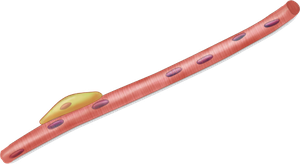
Microscopic Anatomy of Skeletal Muscle
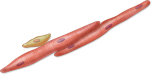
Skeletal muscle fibers are formed by the fusion of myoblasts, resulting in multinucleated cells. Satellite cells are unfused myoblasts that can differentiate if tissue is injured.
Key Point: Muscle fibers are elongated and extend the length of the muscle.
Key Point: Myoblasts contribute nuclei during development.
Cardiac Muscle
Structure and Function
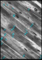
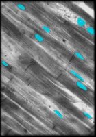
Cardiac muscle is the main muscle of the heart, responsible for pumping blood. Cardiac muscle cells are branched, shorter, and thicker than skeletal muscle fibers, and typically have one or two nuclei.
Key Point: Cardiac muscle cells are joined by intercalated discs, which contain desmosomes and gap junctions.
Key Point: Cardiac muscle is autorhythmic and contracts to its full extent with each heartbeat.
Smooth Muscle
Structure and Function
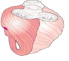
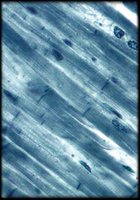
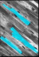
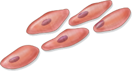
Smooth muscle lines the walls of viscera and is found in longitudinal or circular arrangements. It is responsible for involuntary movements such as peristalsis in the intestine.
Key Point: Smooth muscle cells are spindle-shaped, uni-nucleated, and lack striations.
Key Point: Actin and myosin filaments are present, but the ratio of thin-to-thick filaments is higher than in striated muscle.
Muscle Functions
Major Functions of Muscle Tissue
Muscle tissue is essential for movement, posture, thermogenesis, and stabilization of joints.
Production of Movement: Muscles move body parts, blood, lymph, food, bile, urine, semen, and newborns through the birth canal.
Maintenance of Posture: Muscle contraction allows us to remain upright.
Thermogenesis: Generation of heat via shivering.
Stabilization of Joints: Muscles maintain joint integrity.
Characteristics of Muscle Tissue
Properties of Muscle Tissue
Muscle tissue exhibits several key properties:
Excitability: Ability to receive and respond to stimuli.
Contractility: Ability to shorten forcibly when stimulated.
Conductivity: Electrical change travels along plasma membrane.
Extensibility: Ability to be stretched.
Elasticity: Ability to recoil and resume original length.
Organization of Muscles
Gross Anatomy of Skeletal Muscle
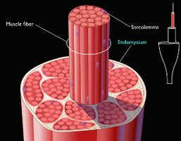
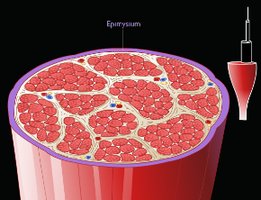
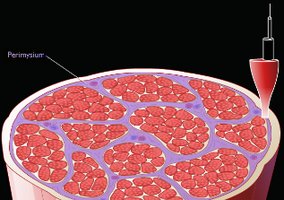
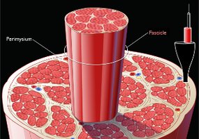
Skeletal muscle is organized into layers of connective tissue: epimysium, perimysium, and endomysium. These layers provide protection, sites for blood vessel and nerve distribution, and attachment to skeleton.
Epimysium: Surrounds the whole muscle.
Perimysium: Surrounds fascicles (bundles of muscle fibers).
Endomysium: Surrounds individual muscle fibers.
Microscopic Anatomy of Skeletal Muscle
Muscle Fiber Structure
Each skeletal muscle cell is known as a muscle fiber. The plasma membrane is called the sarcolemma, and the cytoplasm is called sarcoplasm. Sarcoplasm contains mitochondria, glycogen granules, myofibrils, and sarcoplasmic reticuli.
Key Point: Myofibrils are rodlike structures composed of myofilaments (thick and thin filaments).
Key Point: Sarcolemma has invaginations called transverse tubules (T tubules).
Myofilaments and Sarcomeres
Myofibrils are made up of repeating units called sarcomeres, which are the functional units of muscle contraction.
Thick Filaments: Composed of myosin.
Thin Filaments: Composed of actin, tropomyosin, and troponin.
Sarcomere: Contains regions of thin filaments, thick filaments, and overlapping areas.
Muscle Contraction
Sliding Filament Theory
Muscle contraction occurs when thin filaments slide over thick filaments, shortening the sarcomere and thus the muscle fiber.
Key Point: The process involves excitation, excitation-contraction coupling, contraction, and relaxation.
Neuromuscular Junction and Excitation
The neuromuscular junction is where a motor neuron innervates a muscle fiber. The release of acetylcholine (ACh) from the neuron triggers muscle excitation.
Key Point: ACh binds to receptors on the motor end plate, causing depolarization and initiation of an action potential.
Excitation-Contraction Coupling
The action potential travels along the sarcolemma and T-tubules, triggering the release of Ca2+ from the sarcoplasmic reticulum. Ca2+ binds to troponin, exposing myosin binding sites on actin.
Key Point: This process links muscle stimulation to contraction.
Crossbridge Cycling
Crossbridge cycling involves four steps: crossbridge formation, power stroke, release of myosin head, and reset of myosin head. ATP is required for these steps.
Key Point: The cycle continues as long as Ca2+ and ATP are available.
Muscle Metabolism
Supplying Energy for Muscle Contraction
Muscle fibers generate ATP through three mechanisms:
Immediate Supply: Phosphagen system (creatine phosphate).
Short-Term Supply: Anaerobic cellular respiration (glycolysis and fermentation).
Long-Term Supply: Aerobic cellular respiration (mitochondria).
Skeletal Muscle Fiber Types
Classification of Muscle Fiber Types
Muscle fibers are classified based on contraction speed and ATP supply:
Slow Oxidative (Type I): Slow, fatigue-resistant, aerobic, red appearance.
Fast Oxidative (Type IIa): Intermediate, aerobic, less myoglobin.
Fast Glycolytic (Type IIb): Fast, powerful, anaerobic, white appearance.
Muscle Fatigue and Tension
Muscle Fatigue
Muscle fatigue is the reduced ability to produce tension, often due to decreased glycogen stores, ion imbalances, or impaired crossbridge cycling.
Key Point: Fatigue can result from problems at the neuromuscular junction, excitation-contraction coupling, or crossbridge cycling.
Measurement of Muscle Tension
Muscle tension is measured using a myogram. Muscle twitch consists of latent, contraction, and relaxation periods.
Key Point: Recruitment and changes in stimulus frequency affect muscle tension.
Types of Muscle Contractions
Isometric and Isotonic Contractions
Isometric: Muscle tension increases, but length remains the same (e.g., pushing against a wall).
Isotonic: Muscle tension overcomes resistance, resulting in movement. Includes concentric (muscle shortens) and eccentric (muscle lengthens) contractions.
Smooth Muscle Tissue
Microscopic Anatomy
Smooth muscle cells are fusiform, small, and have a central nucleus. They lack sarcomeres and Z discs, contributing to their smooth appearance.
Key Point: Contractile proteins are arranged between dense bodies and dense plaques.
Key Point: Myosin heads are present along the entire length, allowing latchbridge mechanism.
Smooth Muscle Contraction
Contraction is initiated by Ca2+ binding to calmodulin, activating myosin light-chain kinase, which phosphorylates myosin heads. Relaxation requires removal of Ca2+ and dephosphorylation of myosin.
Key Point: Smooth muscle can maintain contraction without ATP via the latchbridge mechanism.
Functional Categories of Smooth Muscle
Multiunit Smooth Muscle: Contracts individually, found in the eye, skin, airways, arteries.
Single-unit Smooth Muscle: Contracts in unison, found in digestive, urinary, reproductive tracts, blood vessels.
Effects of Exercise and Aging on Muscle
Exercise Effects
Hypertrophy: Increase in muscle size, more mitochondria, glycogen, ATP, and myofibrils.
Hyperplasia: Increase in number of muscle fibers (limited).
Atrophy: Decrease in muscle fiber size and function due to lack of exercise.
Summary Table: Muscle Tissue Types
Type | Control | Structure | Location |
|---|---|---|---|
Skeletal | Voluntary | Striated, multinucleated | Attached to bones |
Cardiac | Involuntary | Striated, branched, 1-2 nuclei | Heart |
Smooth | Involuntary | Non-striated, spindle-shaped, single nucleus | Walls of internal organs |
Additional info: Academic context was added to clarify muscle fiber types, contraction mechanisms, and metabolic pathways for ATP generation. Images were included only when directly relevant to the explanation of the paragraph.
