 Indietro
Comprehensive Study Notes: The Axial and Appendicular Skeleton
Guida di studio - Note intelligenti
 Appunti personalizzati basati sui tuoi materiali, ampliati con definizioni chiave, esempi e contesto.
The Human Skeleton
Overview of the Skeletal System
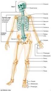
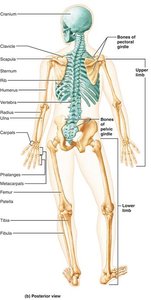
The human skeleton is a structural framework composed of bones, cartilages, joints, and ligaments. It is divided into two major divisions: the axial skeleton and the appendicular skeleton. The skeleton accounts for approximately 20% of body mass and serves critical functions in support, protection, and movement.
Axial Skeleton: Consists of 80 bones, including the skull, vertebral column, and thoracic cage. Forms the longitudinal axis of the body.
Appendicular Skeleton: Composed of bones of the limbs and girdles that attach them to the axial skeleton.
The Axial Skeleton
Skull: Structure and Function
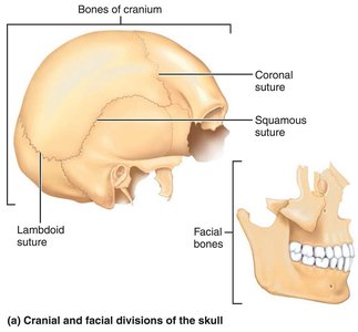
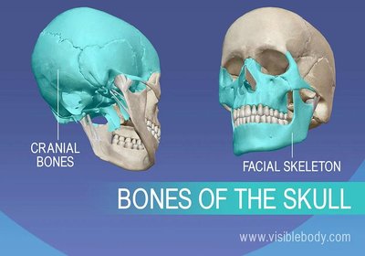
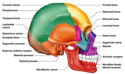
The skull is formed by two sets of bones: cranial bones and facial bones. These bones are mostly flat and firmly locked together by immovable joints called sutures.
Cranial Bones: Enclose and protect the brain, provide attachment sites for head and neck muscles.
Facial Bones: Form the framework of the face, contain cavities for special sense organs, provide openings for air and food passage, secure teeth, and anchor facial muscles.
Skull Geography and Cavities
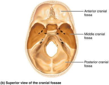
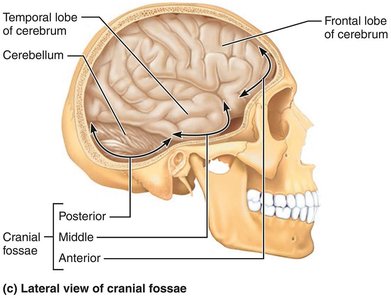
The cranium is divided into a vault (calvaria) and a base. The cranial base is internally divided into three fossae: anterior, middle, and posterior, which house different regions of the brain. The skull also contains other cavities such as the middle and internal ear, nasal cavity, orbits, and sinuses.
Cranial Vault: Forms the superior, lateral, and posterior portion of the skull.
Cranial Base: Forms the inferior aspect and is divided into fossae.
Openings: The skull has 85 named openings (foramina, canals, fissures) for passage of nerves and blood vessels.
Skull Bones and Markings
Foramina: Openings for blood vessels and nerves.
Processes: Projections for muscle attachment.
Sutures: Immovable joints connecting skull bones.
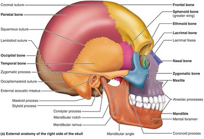
Cranial Bones
The cranium comprises eight cranial bones:
Frontal bone
Parietal bones (left and right)
Occipital bone
Temporal bones (left and right)
Sphenoid bone
Ethmoid bone
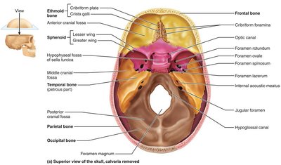
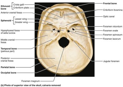
Frontal Bone
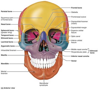
The frontal bone forms the anterior portion of the cranium, including the forehead and superior wall of the orbits. It contains the glabella and frontal sinuses.
Parietal Bones
Two large parietal bones form most of the superior and lateral aspects of the cranial vault. Four major sutures mark their articulations.
Occipital Bone
Forms the posterior wall and base of the skull, contains the foramen magnum, occipital condyles, and sites for muscle attachment. 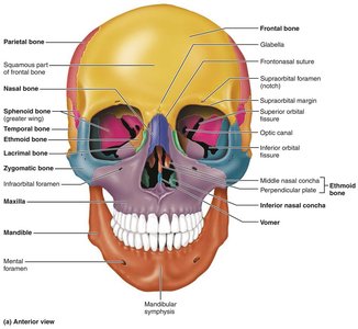
Temporal Bones
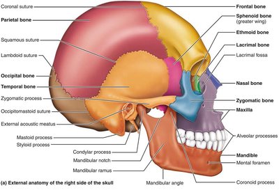
Paired bones making up the inferolateral aspects of the skull and parts of the cranial base. They include squamous, tympanic, and petrous parts, and contain important foramina and processes. 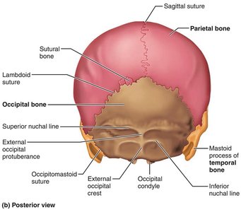
Sphenoid Bone
A complex, bat-shaped bone that articulates with all other cranial bones. Contains sphenoidal sinuses, sella turcica, and several foramina.
Ethmoid Bone
The deepest skull bone, forming the roof of the nasal cavity and part of the medial wall of the orbits. Contains cribriform plates, crista galli, perpendicular plate, and ethmoidal air cells.
Sutural Bones
Tiny, irregularly shaped bones that appear within sutures.
Facial Bones
The facial skeleton is made up of 14 bones, 12 of which are paired. These include the mandible, maxillary bones, zygomatic bones, nasal bones, lacrimal bones, palatine bones, vomer, and inferior nasal conchae. 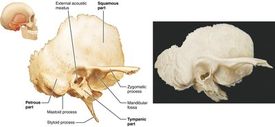
Bony Boundaries of the Orbits and Nasal Cavity
Orbits
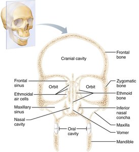
The orbits are cavities that encase the eyes and lacrimal glands, formed by parts of seven bones.
Roof: Frontal bone, sphenoid bone
Lateral Wall: Zygomatic bone, sphenoid bone
Floor: Maxilla, zygomatic bone, palatine bone
Medial Wall: Ethmoid bone, lacrimal bone, maxilla, sphenoid bone
Nasal Cavity
The nasal cavity is formed by several bones and contains paranasal sinuses.
Roof: Nasal bones, frontal bone, ethmoid bone, sphenoid bone
Floor: Maxilla, palatine bone
Lateral Walls: Maxilla, ethmoid bone, inferior nasal conchae, palatine bone
Medial Wall (Nasal Septum): Ethmoid bone (perpendicular plate), vomer
The Vertebral Column
Structure and Regions
The vertebral column extends from the skull to the pelvis and functions to transmit weight, protect the spinal cord, and provide attachment points. It contains 26 irregular bones called vertebrae in five major regions:
Cervical: 7 vertebrae (neck)
Thoracic: 12 vertebrae (upper back)
Lumbar: 5 vertebrae (lower back)
Sacrum: 5 fused vertebrae (pelvis)
Coccyx: 4 fused vertebrae (end of column)
Curvatures
The vertebral column exhibits natural curvatures:
Cervical: Convex anteriorly
Thoracic: Concave anteriorly
Lumbar: Convex anteriorly
Sacral: Concave anteriorly
Functions of Spinal Curvatures and Intervertebral Discs
Spinal Curvatures: Increase resilience and flexibility, help maintain balance and upright posture.
Intervertebral Discs: Cushion vertebrae during movement, allow for bending and twisting.
Common Function: Both contribute to shock absorption and flexibility.
Abnormal Spinal Curvatures
Scoliosis: Abnormal lateral rotation, may lead to breathing difficulties.
Kyphosis: Abnormal dorsal thoracic curvature (hunchback).
Lordosis: Accentuation of lumbar curvature (swayback).
Structure of a Typical Vertebra
Body: Main weight-bearing structure.
Vertebral Arch: Formed by pedicles and laminae.
Processes: Spinous, transverse, superior and inferior articular processes.
Regional Features of Vertebrae
Cervical: Smallest, transverse foramina, bifid spinous processes.
Thoracic: Articular facets for ribs, heart-shaped body, long spinous processes.
Lumbar: Largest, kidney-shaped body, short blunt spinous processes.
The Thoracic Cage
Structure and Function
The thoracic cage forms a protective cage for major organs and is composed of the sternum, ribs, and thoracic vertebrae.
Sternum: Manubrium, body, xiphoid process.
Ribs: True ribs (1-7), false ribs (8-12), floating ribs (11-12).
Functions: Protection, support, respiration.
True vs. False Ribs
True Ribs (1-7): Attach directly to the sternum via costal cartilage.
False Ribs (8-12): Attach indirectly or not at all.
Floating Ribs (11-12): Do not attach to the sternum.
The Appendicular Skeleton
Pectoral (Shoulder) Girdle
Composed of the clavicle and scapula, allowing for free movement of the upper limb.
Clavicle: Collarbone, fractures anteriorly to protect arteries.
Scapula: Shoulder blade, triangular with three borders.
Bones of the Upper Limb
Humerus: Arm bone, articulates with scapula, radius, and ulna.
Ulna: Forms elbow joint, olecranon and coronoid processes.
Radius: Major contribution to wrist joint.
Hand: Carpals (wrist), metacarpals (palm), phalanges (fingers).
Pelvic Girdle
Hip Bones: Ilium, ischium, pubis, fused at acetabulum.
Functions: Supports upper body weight, protects organs.
Bones of the Lower Limb
Femur: Thigh bone, largest and strongest.
Patella: Knee cap, protects knee joint.
Tibia: Medial, weight-bearing.
Fibula: Lateral, does not bear weight.
Foot: Tarsals (ankle), metatarsals (sole), phalanges (toes).
Arches of the Foot
Purpose: Support weight, maintained by bone shapes, ligaments, and tendons.
Types: Medial and lateral longitudinal arches, transverse arch.
Joints
Classification and Function
Joints, or articulations, hold bones together and allow for mobility. They are classified functionally and structurally.
Functional Classification: Synarthroses (immovable), amphiarthroses (slightly movable), diarthroses (freely movable).
Structural Classification: Fibrous, cartilaginous, synovial.
Fibrous Joints
Suture: Only found in skull, immovable.
Syndesmosis: Bones connected by ligaments.
Gomphosis: Peg-in-socket joint (e.g., tooth in socket).
Cartilaginous Joints
Synchondrosis: Hyaline cartilage unites bones, immovable.
Symphysis: Fibrocartilage unites bones, slightly movable (e.g., pubic symphysis, intervertebral joints).
Synovial Joints
Features: Articular cartilage, joint cavity, synovial fluid, fibrous capsule, synovial membrane, ligaments.
Bursae: Flattened sacs lined with synovial membrane.
Tendon Sheath: Elongated bursa around a tendon.
Factors Affecting Synovial Joint Stability
Shape of articular surfaces
Number and positioning of ligaments
Muscle tone
Types of Synovial Joints
Classified by shape and movement.
Inflammatory Conditions Associated with Joints
Bursitis: Inflammation of a bursa.
Tendonitis: Inflammation of tendon sheaths.
Arthritis: Over 100 types, most widespread crippling disease.
Osteoarthritis: Most common, related to aging.
Rheumatoid Arthritis: Autoimmune, leads to deformities.
Bone Health and Clinical Considerations
Importance of Load-Bearing Activities
Stimulates bone deposition and increases bone density.
Reduces risk of osteoporosis and fractures.
Bone Imaging Techniques
X-rays: Detailed images of bone and soft tissues.
MRI: Cross-sectional images for complex fractures.
CT Scans: Detailed bone structure analysis.
Prevention and Treatment of Osteoporosis
Medications: Bisphosphonates, SERMs, hormone therapy.
Prevention: Calcium, vitamin D, weight-bearing exercise, avoid smoking/alcohol.
Risk Factors: Age, genetics, lifestyle, decreased estrogen in postmenopausal women.
Role of Hormones in Bone Health
Estrogen: Promotes bone deposition, inhibits resorption.
Testosterone: Increases bone density.
Parathyroid Hormone (PTH): Regulates calcium, stimulates bone resorption.
Exercise Guidelines for Strong Bones
Weight-bearing exercises, resistance training, flexibility exercises.
Screening for Bone Health
Bone density screening (DEXA scan), risk assessments, early detection.
Role of Rehabilitation in Bone Healing
Occupational therapy, physical therapy, post-fracture rehabilitation.
Summary Table: Axial vs. Appendicular Skeleton
Division | Main Components | Functions |
|---|---|---|
Axial Skeleton | Skull, vertebral column, thoracic cage | Support, protection, attachment for muscles |
Appendicular Skeleton | Pectoral girdle, pelvic girdle, limbs | Movement, manipulation of environment |
Summary Table: Types of Joints
Type | Structural Classification | Functional Classification | Examples |
|---|---|---|---|
Fibrous | Suture, syndesmosis, gomphosis | Synarthroses | Skull sutures, tooth in socket |
Cartilaginous | Synchondrosis, symphysis | Amphiarthroses | Pubic symphysis, intervertebral joints |
Synovial | Joint cavity, synovial fluid | Diarthroses | Knee, shoulder, hip |
Key Equations
Bone Density Calculation:
Mechanical Stress:
Conclusion
The skeletal system is essential for support, protection, movement, and mineral storage. Understanding the structure and function of the axial and appendicular skeleton, as well as the classification and function of joints, is fundamental for students of anatomy and physiology. Regular exercise, proper nutrition, and early screening are crucial for maintaining bone health throughout life.
