 Indietro
Comprehensive Study Notes: The Muscular System (Anatomy & Physiology)
Guida di studio - Note intelligenti
 Appunti personalizzati basati sui tuoi materiali, ampliati con definizioni chiave, esempi e contesto.
The Muscular System
Muscle Attachments and Interactions
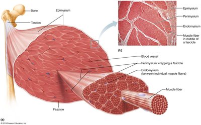
The muscular system is essential for movement, posture, and various bodily functions. Most muscles span joints and are attached to bones at two or more places, typically referred to as the origin (fixed attachment) and insertion (movable attachment). Attachments are often indirect, involving tendons or aponeuroses.
Origin: The stationary attachment point of the muscle.
Insertion: The movable attachment point.
Indirect Attachments: Muscles connect to bones via tendons (cord-like) or aponeuroses (sheet-like).
Arrangement of Fascicles
All skeletal muscles are composed of bundles called fascicles. The arrangement of fascicles determines muscle shape and function.
Parallel: Fascicles run parallel to the long axis (e.g., sartorius).
Fusiform: Spindle-shaped with a central belly (e.g., biceps brachii).
Pennate: Fascicles attach obliquely to a tendon (unipennate, bipennate, multipennate; e.g., rectus femoris, deltoid).
Convergent: Fascicles converge toward a single tendon (e.g., pectoralis major).
Circular: Fascicles arranged in concentric rings, closing openings (e.g., orbicularis oris).
Functional Groups of Muscles
Skeletal muscles work together or in opposition to produce movement. They are classified into functional groups:
Prime Movers (Agonists): Provide the main force for a movement (e.g., biceps brachii for elbow flexion).
Antagonists: Oppose or reverse a movement; help control movement and prevent overshoot (e.g., triceps brachii for elbow extension).
Synergists: Assist prime movers by adding force or reducing undesirable movements (e.g., finger flexors crossing wrist and phalangeal joints).
Fixators: Stabilize the origin of the prime mover (e.g., muscles involved in posture).
Naming Skeletal Muscles
Muscles are named based on several criteria:
Location: Bone or body region (e.g., temporalis, intercostal).
Shape: (e.g., deltoid is triangular, trapezius forms a trapezoid).
Relative Size: Maximus, minimus, longus, brevis (e.g., gluteus maximus).
Direction of Fibers: Rectus (straight), transverse, oblique (e.g., rectus femoris).
Number of Origins: Biceps (two), triceps (three), quadriceps (four).
Location of Attachments: Origin and insertion indicated (e.g., sternocleidomastoid).
Action: Flexor, extensor, adductor (e.g., adductor longus).
Muscles of the Head: Facial Expression
Facial Muscles and Their Functions
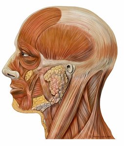
Muscles of facial expression insert into the skin or other muscles, not bones, and are innervated by the facial nerve. They allow for a wide range of expressions and movements.
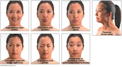
Epicranius: Main muscle of the scalp, with frontal and occipital bellies. The frontal belly raises eyebrows and wrinkles the forehead; the occipital belly fixes the aponeurosis and pulls the scalp posteriorly.
Orbicularis oculi: Surrounds the rim of the orbit; protects eyes by blinking and squinting, draws eyebrows down.
Zygomaticus (major and minor): From zygomatic bone to mouth; responsible for smiling.
Orbicularis oris: Encircles lips; closes, purses, and protrudes lips.
Mentalis: From mandible to chin; protrudes lower lip (pout), wrinkles chin.
Buccinator: From mandible/maxilla to orbicularis oris; whistling, sucking, holds food in place during chewing.
Platysma: From fascia of chest to mandible; depresses mandible, tenses skin of neck.
Muscles of Mastication (Chewing)
Major Muscles Involved in Chewing
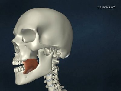
Four pairs of muscles are involved in mastication, all innervated by the mandibular division of cranial nerve V.
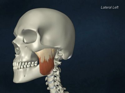
Masseter: Prime mover of jaw closure; originates from zygomatic bone and inserts on mandible.
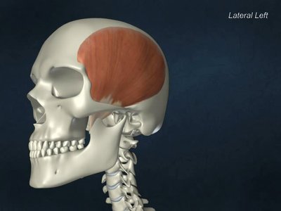
Temporalis: Elevates and retracts mandible; maintains closed jaw at rest.
Medial and Lateral Pterygoids: Provide additional jaw movements, including side-to-side grinding.
Buccinator: Compresses cheek, keeps food between teeth during chewing.
Muscles of the Neck and Vertebral Column
Head and Trunk Movements
Muscles originating from the axial skeleton move the head and trunk. Key muscles include:
Sternocleidomastoid: Prime mover of head flexion; rotates head to opposite side, tilts to same side.
Scalenes: Elevate first two pairs of ribs, assist in neck flexion and rotation.
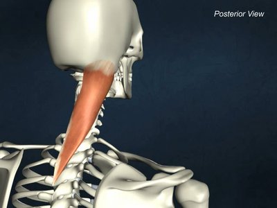
Splenius: Extends or rotates head; superficial, bandage-like muscle.
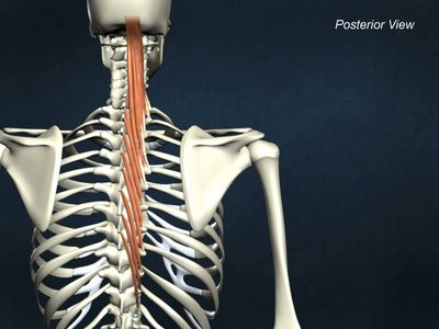
Erector Spinae: Prime mover of back extension; consists of iliocostalis, longissimus, and spinalis columns.
Quadratus Lumborum: Flexes vertebral column laterally, extends lumbar spine, maintains upright posture.
Semispinalis: Deep composite muscle; extends vertebral column and head, synergistic with sternocleidomastoid.
Muscles of Respiration
Thoracic Muscles Involved in Breathing
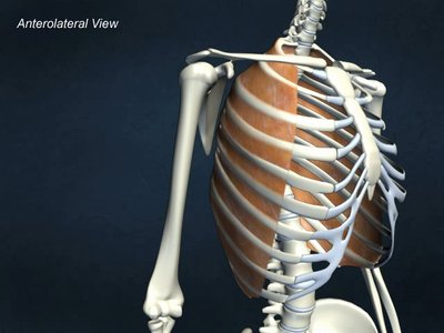
Respiratory muscles alter the size of the thoracic cavity to facilitate breathing.
External Intercostals: Elevate rib cage during inspiration.
Internal Intercostals: Depress rib cage during forced expiration.
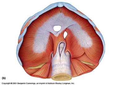
Diaphragm: Dome-shaped; prime mover of inspiration, contracts to increase thoracic volume.
Muscles of the Abdominal Wall
Structure and Function
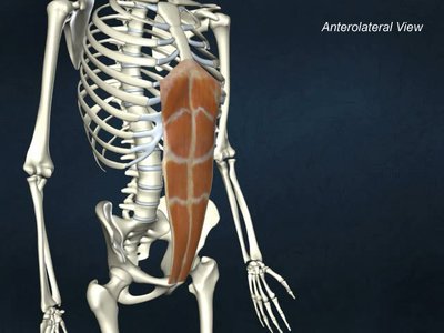
The abdominal wall is formed by four paired muscles, which provide support, movement, and protection for abdominal organs.
Rectus Abdominis: Medial, superficial; flexes vertebral column, stabilizes pelvis.
External Oblique: Largest, most superficial lateral muscle; increases abdominal pressure, flexes vertebral column, rotates trunk.
Internal Oblique: Deeper, similar actions as external oblique.
Transversus Abdominis: Deepest; compresses abdominal contents.
Muscles of the Pelvic Floor and Perineum
Support and Function
These muscles support pelvic organs and control openings of the urethra, vagina, and anus.
Levator Ani: Supports pelvic viscera, forms supportive sling at anorectal junction, resists downward increases in intra-abdominal pressure.
Coccygeus: Supports pelvic viscera, pulls coccyx forward after childbirth.
Urogenital Diaphragm: Supports pelvic organs, constricts urethra.
Superficial Muscles of the Thorax: Arm Movements
Muscles Acting on the Shoulder and Arm
These muscles move the arm and shoulder, providing a wide range of motion.
Pectoralis Major: Prime mover of arm adduction and medial rotation; assists in arm flexion.
Latissimus Dorsi: Broad, flat back muscle; prime mover of arm extension, adduction, and medial rotation.
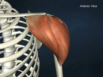
Deltoid: Thick, rounded shoulder muscle; prime mover of arm abduction, site of intramuscular injection.
Trapezius: Stabilizes, raises, retracts, and rotates scapula.
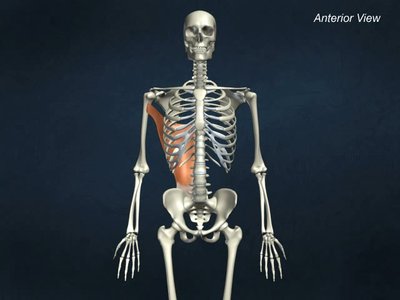
Rhomboids: Deep to trapezius; aid in scapula movements, such as squaring shoulders.
Summary Table: Major Muscles and Their Actions
Muscle | Origin | Insertion | Action |
|---|---|---|---|
Biceps Brachii | Scapula | Radius | Elbow flexion |
Triceps Brachii | Scapula & Humerus | Ulna | Elbow extension |
Deltoid | Clavicle & Scapula | Humerus | Arm abduction |
Masseter | Zygomatic bone | Mandible | Jaw closure |
Rectus Abdominis | Pubis | Rib cage | Flexes vertebral column |
Latissimus Dorsi | Vertebrae | Humerus | Arm extension, adduction |
Additional info: Academic context was added to clarify muscle functions, naming conventions, and anatomical relationships. Images were included only when directly relevant to the explanation of the paragraph.
