 Indietro
Comprehensive Study Notes: The Respiratory System (Anatomy & Physiology)
Guida di studio - Note intelligenti
 Appunti personalizzati basati sui tuoi materiali, ampliati con definizioni chiave, esempi e contesto.
The Respiratory System
Overview and Structural Organization
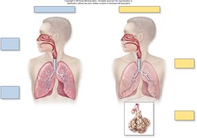
The respiratory system is essential for gas exchange, supplying oxygen to the body and removing carbon dioxide. It is divided into upper and lower respiratory tracts, and further into conducting and respiratory zones.
Upper respiratory tract: Includes the nose, nasal cavity, pharynx, and associated structures.
Lower respiratory tract: Comprises the larynx, trachea, bronchi, bronchioles, alveolar ducts, and alveoli.
Conducting zone: Passages that transport air but do not participate in gas exchange (nose to terminal bronchioles).
Respiratory zone: Structures involved in gas exchange (respiratory bronchioles, alveolar ducts, alveoli).
Respiratory Mucosa and Epithelial Changes
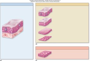
The respiratory tract is lined by a mucosa that varies in thickness and cell type depending on location and function.
Pseudostratified ciliated columnar epithelium: Lines nasal cavity, paranasal sinuses, nasopharynx, trachea, main bronchi.
Simple ciliated columnar epithelium: Found in segmental bronchi and large bronchioles.
Simple ciliated cuboidal epithelium: Lines terminal and respiratory bronchioles.
Simple squamous epithelium: Forms alveolar ducts and alveoli, facilitating gas exchange.
Nonkeratinized stratified squamous epithelium: Protects areas subject to abrasion (oropharynx, laryngopharynx, vocal cords).
Anatomy of the Nasal Cavity and Paranasal Sinuses
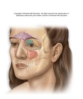
The nasal cavity is divided into regions for olfaction and respiration, and is bordered by bones and cartilage. Paranasal sinuses are air-filled spaces that lighten the skull and aid in voice resonance.
Nasal conchae and meatuses: Increase surface area and turbulence for air filtration.
Paranasal sinuses: Frontal, ethmoidal, sphenoidal, and maxillary sinuses.
Pharynx and Larynx
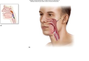
The pharynx is a muscular tube connecting the nasal cavity to the larynx and esophagus, divided into nasopharynx, oropharynx, and laryngopharynx. The larynx is involved in sound production and airway protection.
Nasopharynx: Contains pharyngeal tonsil and auditory tube opening.
Oropharynx: Passage for air and food.
Laryngopharynx: Connects to larynx and esophagus.
Larynx: Supported by cartilages (thyroid, cricoid, arytenoid, etc.), contains vocal folds.
Trachea and Bronchial Tree
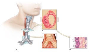
The trachea is a flexible tube supported by C-shaped cartilage rings, leading to the bronchi and branching into smaller airways.
Trachea: Connects larynx to bronchi, lined by pseudostratified ciliated columnar epithelium.
Bronchi: Main, lobar, segmental, and smaller bronchi, supported by cartilage.
Bronchioles: Lack cartilage, have smooth muscle for bronchoconstriction and bronchodilation.
Alveoli and Gas Exchange
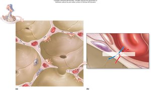
Alveoli are the primary sites for gas exchange, composed of simple squamous epithelium and surrounded by capillaries.
Alveolar type I cells: Form the respiratory membrane for gas diffusion.
Alveolar type II cells: Secrete surfactant to reduce surface tension.
Alveolar macrophages: Remove debris and pathogens.
Respiratory membrane: Consists of alveolar epithelium, fused basement membranes, and capillary endothelium.
Lung Anatomy and Bronchopulmonary Segments
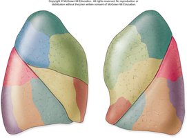
The lungs are divided into lobes and bronchopulmonary segments, each supplied by its own bronchus and blood vessels.
Right lung: Three lobes (superior, middle, inferior).
Left lung: Two lobes (superior, inferior), cardiac notch.
Bronchopulmonary segments: Functional units for localized disease or surgery.
Pleura and Pleural Cavities
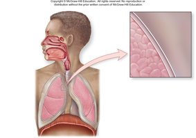
The lungs are enveloped by pleural membranes, creating a pleural cavity that maintains pressure and prevents lung collapse.
Visceral pleura: Covers lung surface.
Parietal pleura: Lines thoracic cavity.
Pleural cavity: Contains serous fluid, reduces friction.
Respiratory Processes and Gas Exchange
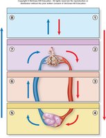
Respiration involves four main processes: pulmonary ventilation, alveolar gas exchange, gas transport, and systemic gas exchange.
Pulmonary ventilation: Movement of air in and out of lungs.
Alveolar gas exchange: Exchange of O2 and CO2 between alveoli and blood.
Gas transport: Movement of gases in blood.
Systemic gas exchange: Exchange of gases between blood and tissues.
Mechanics of Breathing
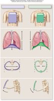
Breathing is driven by changes in thoracic cavity volume and pressure, governed by Boyle's law.
Inspiration: Diaphragm contracts, thoracic volume increases, pressure decreases, air flows in.
Expiration: Diaphragm relaxes, thoracic volume decreases, pressure increases, air flows out.
Boyle's law: (Pressure and volume are inversely related).
Control of Breathing
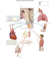
Breathing is regulated by the respiratory centers in the brainstem, responding to chemoreceptors, baroreceptors, and other sensory inputs.
Medullary respiratory center: Controls rhythm and depth of breathing.
Pontine respiratory center: Modifies breathing patterns.
Chemoreceptors: Detect changes in CO2, O2, and pH.
Respiratory Volumes and Capacities
Lung volumes and capacities are important for assessing respiratory health.
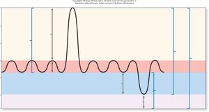
Tidal volume (TV): Air moved per quiet breath (500 mL).
Inspiratory reserve volume (IRV): Max air inhaled after normal inspiration.
Expiratory reserve volume (ERV): Max air exhaled after normal expiration.
Residual volume (RV): Air remaining after forced expiration.
Vital capacity:
Total lung capacity:
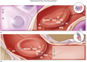
Gas Transport and Exchange
Oxygen and carbon dioxide are transported in the blood by different mechanisms.
Oxygen: Mostly bound to hemoglobin; small amount dissolved in plasma.
Carbon dioxide: Transported as bicarbonate (HCO3-), bound to hemoglobin, or dissolved in plasma.
Chloride shift: Exchange of Cl- and HCO3- in erythrocytes.
Bohr effect: Increased H+ promotes O2 release from hemoglobin.
Haldane effect: Increased O2 release promotes CO2 binding to hemoglobin.
Factors Affecting Gas Exchange
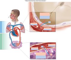
Gas exchange efficiency depends on partial pressure gradients, membrane thickness, surface area, and ventilation-perfusion coupling.
Partial pressure gradients: Drive diffusion of O2 and CO2.
Ventilation-perfusion coupling: Matches airflow and blood flow for optimal exchange.
Membrane thickness: Increased thickness impairs exchange.
Homeostatic Imbalances
Various conditions can disrupt respiratory function, affecting gas exchange and transport.
System | Physiologic Consequences | Clinical Example |
|---|---|---|
Respiratory | Decreased airflow, gas exchange | Asthma, bronchitis |
Skeletal | Impaired thoracic volume changes | Arthritis, deformities |
Muscular | Impaired muscle function | Polio, muscular dystrophy |
Nervous | Decreased stimulation of breathing | Brainstem injury, trauma |
Cardiovascular | Impeded blood flow, decreased gas transport | Pulmonary embolism, anemia |
Summary of Oxygen and Carbon Dioxide Transport
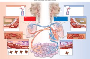
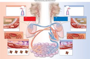
Oxygen: Diffuses from alveoli to blood, transported by hemoglobin, released to tissues.
Carbon dioxide: Produced by tissues, transported as bicarbonate, released into alveoli.
Additional info:
Ventilation-perfusion coupling ensures that areas of the lung receiving air also receive blood flow for efficient gas exchange.
Respiratory volumes and capacities are measured using spirometry for clinical assessment.
