 Indietro
Diabetes Mellitus: Anatomy, Physiology, and Pathophysiology of the Pancreas
Guida di studio - Note intelligenti
 Appunti personalizzati basati sui tuoi materiali, ampliati con definizioni chiave, esempi e contesto.
The Pancreas: Anatomy and Physiology
Location and Structure
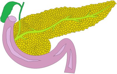
The pancreas is a long, flat organ situated within the abdomen, posterior to the stomach. It measures approximately 12–20 cm in length and possesses both endocrine and exocrine functions.
Exocrine function: Secretes digestive enzymes into the duodenum for carbohydrate, lipid, and protein breakdown.
Endocrine function: Produces hormones, primarily insulin and glucagon, which regulate blood glucose levels.
Cell Types in the Islets of Langerhans
The endocrine portion of the pancreas consists of the islets of Langerhans, which contain five key cell types:
Alpha cells: Produce glucagon, raising blood glucose by triggering glycogen breakdown in the liver.
Beta cells: Produce insulin, lowering blood glucose by promoting glycogen formation and glucose uptake by muscle and fat cells.
Delta cells: Produce somatostatin, which inhibits insulin and glucagon secretion.
Gamma (PP) cells: Produce pancreatic polypeptide, regulating pancreatic function and appetite.
Epsilon cells: Produce ghrelin, inducing hunger and inhibiting insulin secretion.
Blood Glucose Regulation
Hormonal Control and Homeostasis
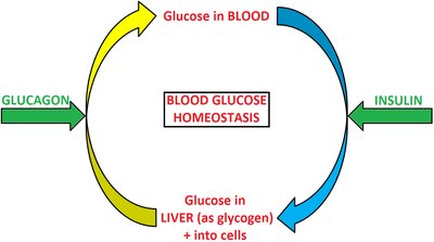
Blood glucose homeostasis is maintained by the antagonistic actions of insulin and glucagon. The normal adult fasting range is 3.5–5.5 mmol/L.
Insulin: Activates glucose transport into liver, fat, and muscle cells, lowering blood glucose.
Glucagon: Activates breakdown of glycogen in the liver, raising blood glucose.
Mechanism of Insulin Action
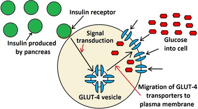
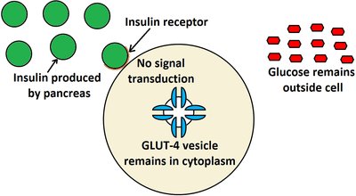
Insulin binds to receptors on cell surfaces, causing glucose transporter proteins (GLUT-4) to move to the plasma membrane, allowing glucose to enter cells via facilitated diffusion.
When blood glucose rises, beta cells release insulin.
Insulin stimulates glucose uptake by cells, decreasing blood glucose.
Diabetes Mellitus: Types and Pathophysiology
Type I Diabetes Mellitus
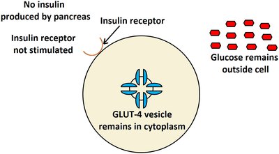
Type I diabetes is an autoimmune disease characterized by destruction of beta cells, resulting in little or no insulin production. It often presents suddenly when most beta cells are lost.
Autoimmune destruction: Lymphocytes and macrophages attack beta cells.
Triggers: Genetic and environmental factors.
Pathophysiology: Over-secretion of glucagon, severe hyperglycaemia, increased fat breakdown, and ketoacidosis.
Rare causes: Pancreatitis or idiopathic origins.
Type II Diabetes Mellitus
Type II diabetes is characterized by insulin resistance. The pancreas still produces insulin, but cells become less responsive. It develops gradually and is linked to environmental and genetic factors.
Triggers: Overweight, age, ethnicity, hypertension, lack of physical activity.
Pathophysiology: Increased insulin production, eventual beta cell exhaustion, reduced insulin receptors.
Reversibility: Type II can be reversed with lifestyle changes; Type I cannot.
Progression: Can take years, with a pre-diabetic state called metabolic syndrome.
Metabolic Syndrome and Obesity
Metabolic Syndrome Criteria
Metabolic syndrome is diagnosed if three or more of the following traits are present:
Trait | Measurement |
|---|---|
Increased waist circumference | male > 40 inches, female > 35 inches |
Increased plasma triglycerides | ≥ 1.71 mmol/L |
Reduced plasma HDL:cholesterol ratio | male < 1.04 mmol/L, female < 1.3 mmol/L |
Elevated blood pressure | ≥ 130/85 mmHg |
Elevated fasting plasma glucose | 5.6–6.9 mmol/L |
Obesity and Type II Diabetes
Obesity contributes to type II diabetes through several mechanisms:
Adipokines: Inflammatory hormones from fat cells decrease insulin sensitivity.
Elevated blood fats: Triglycerides and cholesterol interfere with insulin signaling.
Inflammatory cytokines: Produced by intra-abdominal fat, toxic to beta cells.
Duration of overweight: Prolonged hyperinsulinemia leads to beta cell exhaustion.
Symptoms of Diabetes Mellitus
Key Definitions
Polyuria: Excessive urine production (>3000 mL/day).
Polydipsia: Excessive thirst.
Polyphagia: Excessive hunger.
Paraesthesia: Abnormal sensations (tingling, pins and needles).
Type I Diabetes Symptoms
Polyuria: Glucose in urine acts as a diuretic, causing dehydration.
Polydipsia: Dehydration triggers thirst.
Polyphagia: Reduced glucose metabolism causes hunger.
Weight loss: Fat stores metabolized for energy.
Fatigue: Low energy due to impaired glucose metabolism.
Visual disturbance: Osmotic changes and retinal damage.
Paraesthesia: Nerve dysfunction from hyperglycaemia.
Type II Diabetes Symptoms
Polyuria
Nocturia (nighttime urination)
Polydipsia
Polyphagia
Fatigue
Pruritus (itchy skin)
Recurrent infections (e.g., wounds that don’t heal)
Visual changes
Neuropathy
Diagnosis of Diabetes Mellitus
Diagnostic Tests and Criteria
Diabetes is diagnosed using several laboratory tests:
Test | Normal | Diabetes Mellitus |
|---|---|---|
Glycosylated haemoglobin (HbA1c) | 18–46 mmol/mol | > 48 mmol/mol |
Fasting Plasma Glucose (FPG) | < 7.8 mmol/L | ≥ 7.8 mmol/L |
Oral Glucose Tolerance Test (OGTT) | < 7.8 mmol/L | ≥ 11.1 mmol/L |
Random Plasma Glucose | 4.2–6.4 mmol/L | ≥ 11.1 mmol/L |
HbA1c reflects average blood glucose over ~3 months, useful for long-term monitoring.
Complications of Diabetes Mellitus
Acute Complications
Diabetic ketoacidosis: Most common in type I, caused by insulin deficiency, leading to hyperglycaemia, fat metabolism, acidic ketones, and low blood pH.
Severe hypoglycaemia (insulin shock): Plasma glucose < 3.33 mmol/L, caused by excess insulin or medication, resulting in pallor, tremor, anxiety, seizures, coma.
Chronic Complications
Cardiovascular: Atherosclerosis, hypertension, hypercoagulability, heart failure, poor wound healing.
Neuropathy: Peripheral nerve dysfunction and degeneration.
Nephropathy: Glomerular damage, renal failure (leading cause of dialysis).
Retinopathy: Cataracts, macular degeneration.
Infections: Impaired immunity due to abnormal blood sugar and fat levels.
