 Indietro
Digestive System Anatomy & Physiology: Structured Study Notes
Guida di studio - Note intelligenti
 Appunti personalizzati basati sui tuoi materiali, ampliati con definizioni chiave, esempi e contesto.
Digestive System Overview
Tube Within a Tube Concept
The digestive system is structured as a "tube within a tube," where the gastrointestinal (GI) tract runs from the mouth to the anus. Food is not considered "inside the body" until it is digested and absorbed into the bloodstream, making nutrients accessible to cells.
GI Tract Synonyms: Digestive tract, alimentary canal
Function: Digestion, absorption, and elimination of food
Major Organs of the Digestive Tract
The digestive tract consists of several major organs, each with specialized functions in the breakdown and absorption of nutrients.
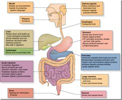
Digestive Processes
1. Ingestion
Ingestion is the intake of food and liquid via the mouth, marking the entry point into the digestive tract.
Location: Mouth
Function: Entry of food and drink
2. Motility
Motility refers to the movement of food and liquid through the GI tract, facilitated by processes such as swallowing, peristalsis, and segmentation.
Swallowing: Voluntary movement of food from mouth to pharynx
Peristalsis: Wave-like contractions propelling food forward
Segmentation: Rhythmic contractions mixing food
3. Secretion
Secretion involves the release of substances by glands into ducts (e.g., saliva) or into the blood (e.g., GI hormones).
Saliva: Contains enzymes, mucin, lysozyme
GI Hormones: Regulate digestive activity
4. Digestion
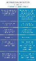
Digestion is the breakdown of food into smaller components, occurring through mechanical and chemical processes.
Mechanical Digestion: Physical breakdown (chewing, churning, segmentation)
Chemical Digestion: Enzymatic breakdown (carbs to monosaccharides, proteins to amino acids, fats to monoglycerides and fatty acids, nucleic acids to nucleotides)
5. Absorption
Absorption is the process by which nutrients cross the epithelial layer of the GI tract and enter the blood or lymph.
Amino acids & monosaccharides: Absorbed into blood
Fatty acids & monoglycerides: Absorbed into lymphatic capillaries
6. Elimination (Defecation)
Elimination is the removal of waste from the body via the anus in the form of feces.
Waste: Material not absorbed as nutrients
Digestive Tract Membranes
Peritoneum
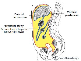
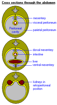
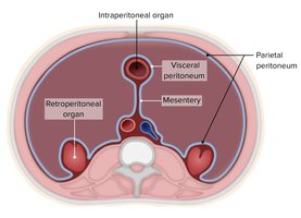
The peritoneum is the largest and most complex serous membrane in the body, consisting of two layers:
Parietal peritoneum: Lines the abdominopelvic cavity
Visceral peritoneum: Wraps around intraperitoneal organs
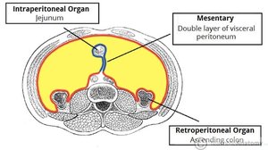
Retroperitoneal organs: Located outside the peritoneum
Main function: Reduces friction between organs
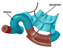
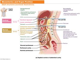
Mesentery
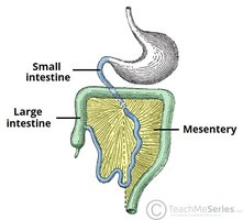
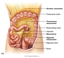
The mesentery is a double layer of visceral peritoneum that anchors organs to the abdominal wall, stores fat, and provides routes for blood vessels, lymphatics, and nerves.
Dorsal mesenteries: Attach to posterior abdominal wall
Ventral mesenteries: Attach to anterior abdominal wall
Lesser and Greater Omentum
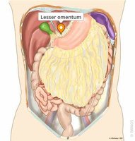
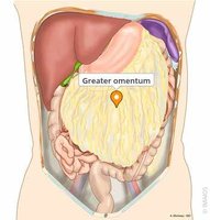
The lesser omentum connects the liver to the lesser curvature of the stomach, while the greater omentum is a large fatty apron that drapes over the intestines.
Mesentery Proper and Mesocolon
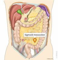
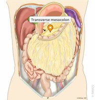
The mesentery proper surrounds the intestines and anchors them to the posterior abdominal wall. The mesocolon anchors sections of the colon.
Digestive Tract Wall Structure
GI Wall Layers
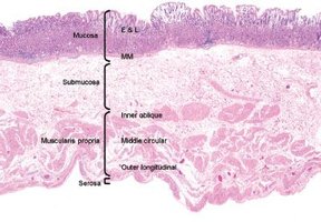
The GI wall from esophagus to large intestine consists of four layers:
Tunica mucosa: Innermost layer, contact with food; includes epithelium, lamina propria, muscularis mucosa
Tunica submucosa: Dense connective tissue, contains blood and lymphatic vessels, glands
Tunica muscularis externa: Two layers of smooth muscle (inner circular, outer longitudinal)
Tunica serosa/adventitia: Serosa (visceral peritoneum) for organs in abdominal cavity; adventitia (connective tissue) for organs outside
Retroperitoneal Organs
Retroperitoneal organs are covered by peritoneum only on their anterior surface and are not suspended by mesentery. They have both tunica adventitia and tunica serosa.
Changes in GI Wall Epithelium
The epithelium of the tunica mucosa changes along the GI tract:
Stratified squamous epithelium: Oral cavity, pharynx, esophagus, distal anal canal (resists abrasion)
Simple columnar epithelium: Stomach, small intestine, large intestine, proximal anal canal (secretion/absorption)
Gastroesophageal Junction
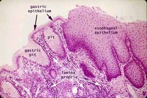
The transition from esophagus to stomach involves a shift from stratified squamous to simple columnar epithelium, forming gastric pits that secrete alkaline mucus.
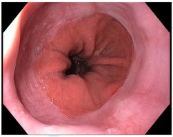
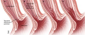
Clinical Connection: GERD and Barrett's Esophagus
The gastroesophageal junction is a high-stress zone. Chronic acid reflux (GERD) can cause Barrett's Esophagus, where columnar cells invade the esophagus, increasing cancer risk.
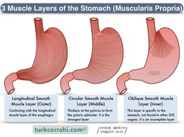
Stomach Muscularis Externa
The stomach has three layers of smooth muscle (inner oblique, middle circular, outer longitudinal) for churning food into chyme, unlike the esophagus which only needs to push food down.
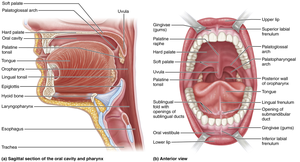
Mouth and Associated Structures
Mouth Anatomy
The mouth is bounded by lips (anterior), fauces (posterior), palate (superior), tongue (inferior), and cheeks (lateral). It is lined with nonkeratinized stratified squamous epithelium.
Salivary Glands and Saliva
Three sets of salivary glands (parotid, sublingual, submandibular) produce saliva, which contains water, ions, buffers, lysozyme, mucin, and salivary amylase.
Salivary amylase: Enzyme for starch digestion
Lysozyme: Antimicrobial
Mucin: Moisture and lubrication
Ions and buffers: Oral pH balance
Teeth
Teeth are responsible for mastication (chewing), a form of mechanical digestion. Enamel covers the crown, and dentin forms the bulk beneath. Periodontal ligaments hold teeth in alveoli of maxilla and mandible.
Tongue
The tongue is a large muscle secured by the lingual frenulum. It repositions food, mixes it with saliva, forms a bolus, and initiates swallowing.
Pharynx and Esophagus
Pharynx
The oropharynx and laryngopharynx serve as common passageways for food, fluids, and air. The nasopharynx has no digestive role. The pharynx is lined with stratified squamous epithelium and encircled by pharyngeal constrictor muscles.
Esophagus
The esophagus is a muscular tube posterior to the trachea, collapsed when not in use. It carries the bolus through the mediastinum and diaphragm to the stomach. The esophageal hiatus is the opening in the diaphragm for the esophagus.
Muscles of the Esophagus
The muscularis externa of the esophagus varies:
Superior third: Striated, skeletal muscle (voluntary)
Middle third: Mixed skeletal and smooth muscle
Inferior third: Smooth muscle (involuntary)
Three Phases of Swallowing (Deglutition)
Swallowing consists of oral, pharyngeal, and esophageal phases, involving coordinated muscle contractions to move food from mouth to stomach.
Stomach Anatomy and Function
Stomach Regions
The stomach has four regions: cardia (near gastroesophageal sphincter), fundus (dome-shaped, holds gas), body (main digestion area), and pylorus (exits to duodenum).
Rugae
Rugae are large folds in the stomach wall that allow stretching during filling. They flatten as the stomach fills, most prominent in fundus and body.
Stomach Mucosa: Gastric Pits and Glands
Gastric pits are indentations in the mucosa opening into gastric glands, which produce gastric juice. Both are lined with simple columnar epithelium.
Gastric Gland Cell Types
Mucous neck cells: Produce mucus
Parietal cells: Produce hydrochloric acid (HCl)
Chief cells: Produce pepsinogen (inactive enzyme)
Enteroendocrine cells: Produce digestive hormones
Pepsin and Protein Digestion
Pepsinogen is converted to pepsin by HCl in the stomach, which digests proteins. HCl first denatures proteins, then pepsin breaks them into peptides. Further breakdown occurs in the small intestine.
Digestive Processes of the Stomach
Motility: Peristalsis
Physical digestion: Churning with three muscle layers
Chemical digestion: Protein digestion begins (pepsin)
Absorption: Limited; alcohol and aspirin absorbed
Chyme
Chyme is the thick, semi-fluid mixture produced by the stomach, which exits via the pyloric sphincter into the small intestine, where most digestion and absorption occur.
Small Intestine Anatomy and Function
Three Regions of Small Intestine
Duodenum: Chemical digestion, neutralizes stomach acid
Jejunum: Bulk absorption of nutrients
Ileum: Absorption of vitamin B12, bile salts, remaining nutrients
Motility: Peristalsis vs Segmentation
Peristalsis propels food forward, while segmentation mixes food and maximizes contact with the lumen wall for absorption.
Special Features of Small Intestine Mucosa
Plicae circularis: Permanent circular folds increasing surface area and slowing chyme movement
Villi: Finger-like projections lined with simple columnar epithelium
Microvilli: Increase surface area further
Goblet cells: Secrete mucin for mucus production
Absorption in Small Intestine
The lamina propria of each villus contains a capillary bed (for sugars and amino acids) and a lacteal (for fats).
Large Intestine Anatomy and Function
Four Regions of Large Intestine
Cecum: Entry point from ileum, appendix attached
Colon: Ascending, transverse, descending, sigmoid
Rectum: Feces storage, rectal valves
Anus: Internal (involuntary) and external (voluntary) sphincters
Special Features of Large Intestine
Taeniae coli: Three longitudinal muscle bands
Haustra: Pocket-like sacs formed by taeniae coli
Functions of Large Intestine
Houses gut microbes: Digestion, vitamin production
Reabsorbs water and electrolytes
Compacts chyme into feces
Absorbs vitamins (e.g., Vitamin K)
Stores feces before defecation
Mucosa of Large Intestine
No villi
Many intestinal glands and goblet cells
Epithelium: Simple columnar, switches to stratified squamous in distal anal canal
Clinical Connection: C. diff Infection
Clostridioides difficile is a severe hospital-acquired infection, often following broad-spectrum antibiotics. It disrupts the healthy microbiome and is resistant to many treatments. Fecal Microbiota Transplants (FMT) are a potential therapy.
Accessory Organs of the Small Intestine
Liver, Gallbladder, and Pancreas
These organs are essential for digestion and absorption in the small intestine.
Liver: Regulates blood composition, breaks down old RBCs, produces bile (bilirubin and bile salts)
Gallbladder: Stores and concentrates bile, releases it during fat digestion
Pancreas: Produces pancreatic juice (digestive enzymes, bicarbonate), regulates blood sugar (insulin, glucagon)
Hepatopancreatic Ampulla and Sphincter
The bile duct and main pancreatic duct fuse at the hepatopancreatic ampulla in the duodenum, controlled by the hepatopancreatic sphincter.
Bile and Pancreatic Juice
Bile: Produced in liver, stored in gallbladder, emulsifies fats
Pancreatic juice: Contains water, bicarbonate (neutralizes acid), digestive enzymes (amylase, proteases, lipases)
Pancreas Dual Function
Endocrine: Regulates blood sugar (insulin, glucagon)
Exocrine: Produces digestive enzymes and bicarbonate
Additional info: Academic context and expanded explanations were added to ensure completeness and clarity for exam preparation.
