 Indietro
Digestive System and Circulatory Pathways: Structure and Function
Guida di studio - Note intelligenti
 Appunti personalizzati basati sui tuoi materiali, ampliati con definizioni chiave, esempi e contesto.
Digestive System and Circulatory Pathways
Overview
This study guide covers the structure and function of the digestive system and the circulatory pathways, focusing on the anatomy, histology, and physiological processes relevant to human Anatomy & Physiology II. Key topics include the pathway of blood through the heart and body, the anatomy of the digestive tract, and the histological organization of digestive organs.
Pathway of Blood Through the Heart and Body
Systemic and Pulmonary Circuits
The human circulatory system is divided into two main circuits: the pulmonary circuit (which carries blood between the heart and lungs) and the systemic circuit (which carries blood between the heart and the rest of the body).
Pulmonary Circuit: Right ventricle → Pulmonary arteries → Lungs → Pulmonary veins → Left atrium
Systemic Circuit: Left ventricle → Aorta → Body tissues → Vena cava → Right atrium
Oxygen-poor blood is pumped from the right side of the heart to the lungs, where it is oxygenated, then returns to the left side of the heart to be pumped throughout the body.
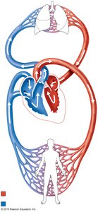
Simplified Representation of Blood Flow
For easier visualization, the pathway of blood can be represented in a simplified schematic, emphasizing the cyclical nature of circulation and the separation of oxygenated and deoxygenated blood.
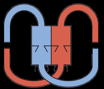
Detailed Pathway of Blood
Right atrium
Tricuspid valve
Right ventricle
Pulmonary semilunar valve
Pulmonary trunk (L/R pulmonary arteries)
Lungs (capillaries surrounding air sacs)
Pulmonary veins
Left atrium
Bicuspid (mitral) valve
Left ventricle
Aortic semilunar valve
Aorta
Systemic capillaries (throughout body)
Vena cava (superior and inferior)
This cycle repeats continuously to maintain oxygen and nutrient delivery to tissues and removal of waste products.
The Digestive System: Anatomy and Function
Major Organs of the Digestive System
The digestive system consists of the alimentary canal (gastrointestinal tract) and accessory organs. The main organs include:
Mouth (oral cavity)
Pharynx
Esophagus
Stomach
Small intestine (duodenum, jejunum, ileum)
Large intestine (cecum, colon, rectum, anal canal)
Anus
Accessory organs include the salivary glands, liver, gallbladder, and pancreas.
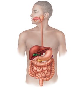
Accessory Digestive Organs
Salivary glands: Parotid, sublingual, and submandibular glands produce saliva to initiate digestion.
Liver: Produces bile for fat emulsification.
Gallbladder: Stores and concentrates bile.
Pancreas: Secretes digestive enzymes and bicarbonate into the small intestine.
Histological Structure of the Alimentary Canal
General Wall Structure
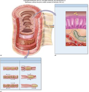
The wall of the alimentary canal consists of four main layers (tunics):
Mucosa: Innermost layer; consists of epithelium, lamina propria, and muscularis mucosae. Functions in secretion, absorption, and protection.
Submucosa: Dense connective tissue containing blood vessels, lymphatics, and nerves (submucosal plexus).
Muscularis externa: Two layers of smooth muscle (inner circular and outer longitudinal) responsible for peristalsis and segmentation. Contains the myenteric nerve plexus.
Serosa (or adventitia): Outermost layer; serosa is a visceral peritoneum, while adventitia is connective tissue in regions outside the peritoneal cavity.
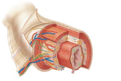
Key Terms for Histology
Lumen: The hollow passageway through which food travels.
Mucosa-associated lymphoid tissue (MALT): Immune tissue found in the mucosa.
Mesentery: Double layer of peritoneum that supports the intestines.
Motility and Absorption in the GI Tract
Types of Motility
The GI tract exhibits two primary types of motility:
Mixing (segmentation): Local contractions that mix food with digestive juices and increase contact with the mucosa for absorption.
Propulsion (peristalsis): Coordinated contractions that move food along the tract.
Absorption
Absorption is the process by which digested nutrients pass from the lumen of the GI tract through the epithelial cells into the blood or lymphatic capillaries.
Most nutrients (carbohydrates, amino acids, water-soluble vitamins) are absorbed into blood capillaries.
Lipids and fat-soluble vitamins are absorbed into lymphatic capillaries (lacteals).

Oral Cavity and Pharynx
Anatomy of the Oral Cavity and Pharynx
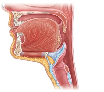
The oral cavity and pharynx are the entry points for food and air. Key structures include:
Hard palate
Soft palate
Uvula
Tongue
Oropharynx
Laryngopharynx
Epiglottis
Esophagus
Trachea
Histology of the Esophagus
Layers of the Esophagus
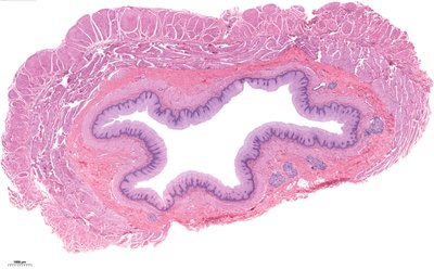
The esophagus is a muscular tube lined with non-keratinized stratified squamous epithelium, which protects against abrasion from food particles. Key histological features include:
Mucosa: Non-keratinized stratified squamous epithelium, lamina propria (loose connective tissue), muscularis mucosae (smooth muscle)
Submucosa: Contains esophageal (mucous) glands for lubrication
Muscularis externa: Skeletal muscle in the upper third, smooth muscle in the lower two-thirds
Anatomy and Histology of the Stomach
Gross Anatomy of the Stomach
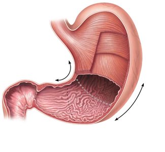
The stomach is divided into four main regions: cardia, fundus, body, and pylorus. It has two curvatures (greater and lesser) and is bounded by the cardiac and pyloric sphincters.
Rugae: Folds in the mucosa that allow for expansion
Muscularis externa: Three layers—longitudinal, circular, and oblique—for churning food
Histological Layers of the Stomach Wall
Mucosa: Contains gastric pits and glands
Submucosa
Muscularis externa: Oblique, circular, and longitudinal muscle layers
Serosa
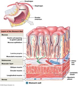
Gastric Glands and Cell Types
Surface mucous cells: Secrete alkaline mucus
Mucous neck cells: Secrete less alkaline mucus
Parietal cells: Secrete hydrochloric acid (HCl) and intrinsic factor
Chief cells: Secrete pepsinogen and gastric lipase
G-cells (enteroendocrine cells): Secrete hormones such as gastrin
Small Intestine: Structure and Function
Regions of the Small Intestine
Duodenum: Receives chyme from the stomach and digestive secretions from the pancreas and liver. Contains Brunner’s glands in the submucosa.
Jejunum: Main site for nutrient absorption; tall, slender villi.
Ileum: Contains Peyer’s patches (lymphoid tissue); villi are shorter and broader.
Histological Features
Circular folds (plicae circulares): Increase surface area for absorption
Villi: Finger-like projections of the mucosa
Microvilli: Microscopic extensions of epithelial cells forming the brush border
Goblet cells: Secrete mucus
Lacteals: Lymphatic capillaries in villi for lipid absorption
Large Intestine: Structure and Function
Regions of the Large Intestine
Cecum (with appendix)
Ascending colon
Transverse colon
Descending colon
Sigmoid colon
Rectum
Anal canal
Key features include the ileocecal valve, haustra (pouches), teniae coli (longitudinal muscle bands), and abundant goblet cells for mucus secretion.
Liver and Pancreas: Structure and Function
Liver Anatomy and Histology
Lobes: Right and left
Hepatic lobule: Functional unit of the liver, hexagonal in shape
Portal triad: Bile duct, branch of hepatic portal vein, branch of hepatic artery
Central vein: Drains blood from the lobule
Hepatocytes: Liver cells that process nutrients and detoxify blood
Hepatic sinusoids: Capillary spaces for blood flow and exchange
Pancreas
Produces digestive enzymes and bicarbonate
Delivers secretions to the duodenum via the pancreatic duct
Summary Table: Layers of the Alimentary Canal
Layer | Main Components | Function |
|---|---|---|
Mucosa | Epithelium, lamina propria, muscularis mucosae | Secretion, absorption, protection |
Submucosa | Connective tissue, blood vessels, nerves | Support, nourishment, innervation |
Muscularis externa | Inner circular and outer longitudinal muscle | Motility (peristalsis, segmentation) |
Serosa/Adventitia | Connective tissue, mesothelium (serosa only) | Protection, structural support |
Additional info: For further study, refer to histology guides and laboratory slides to identify specific cell types and tissue structures in the digestive tract. Understanding the relationship between structure and function is essential for mastering Anatomy & Physiology.
