 Indietro
Digestive System: Liver, Gallbladder, Pancreas, and Intestines
Guida di studio - Note intelligenti
 Appunti personalizzati basati sui tuoi materiali, ampliati con definizioni chiave, esempi e contesto.
Liver, Gallbladder, and Pancreas
Overview
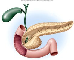
The liver, gallbladder, and pancreas are essential organs that release secretions into the small intestine to facilitate digestion. Their coordinated functions ensure the breakdown and absorption of nutrients.
Liver: Produces bile, metabolizes nutrients, detoxifies blood.
Gallbladder: Stores and concentrates bile.
Pancreas: Secretes digestive enzymes and hormones.
Functions of the Liver
Metabolic and Regulatory Roles
The liver performs numerous vital functions related to metabolism, detoxification, and storage.
Carbohydrate metabolism: Maintains blood glucose levels via glycogen storage and gluconeogenesis.
Fat metabolism: Synthesizes cholesterol and lipoproteins; breaks down fatty acids.
Protein metabolism: Deaminates amino acids, produces plasma proteins.
Removal of drugs and hormones: Detoxifies blood.
Excretion and synthesis of bile: Essential for fat digestion.
Storage: Stores vitamins (A, D, B12), minerals (iron), and glycogen.
Phagocytosis: Kupffer cells remove old RBCs and WBCs.
Removal of toxins: Processes alcohol, ammonia, and other toxins.
Gross Anatomy of the Liver
Lobes and Ligaments
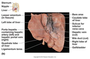
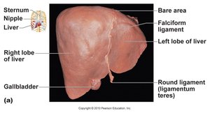
The liver is a large, reddish-brown organ located inferior to the diaphragm. It is divided into four lobes and is supported by several ligaments.
Right lobe and left lobe separated by the falciform ligament.
Caudate lobe and quadrate lobe are smaller lobes.
Round ligament (ligamentum teres): Remnant of the fetal umbilical vein.
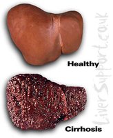
Liver Cirrhosis
Pathology and Progression
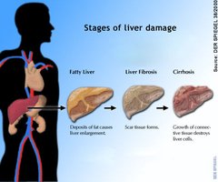
Cirrhosis is a chronic liver disease characterized by progressive destruction of liver tissue and replacement with fibrous scar tissue.
Fatty liver: Initial stage with fat accumulation.
Liver fibrosis: Formation of scar tissue.
Cirrhosis: Extensive fibrosis destroys liver cells and impairs function.
Hepatitis
Types and Transmission
Hepatitis is inflammation of the liver, often caused by viral infections. Different types have distinct modes of transmission and clinical outcomes.
Hepatitis A: Acute diarrheal illness, transmitted via contaminated food/water.
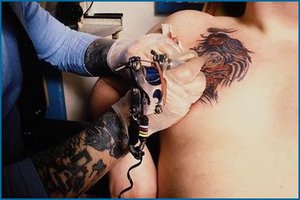
Hepatitis B: Transmitted via blood/body fluids; vaccine available.
Hepatitis C: Chronic, blood/body fluid transmission; increased risk of liver cancer.
Hepatic Circulation
Arterial and Venous Blood Flow
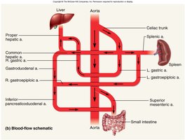
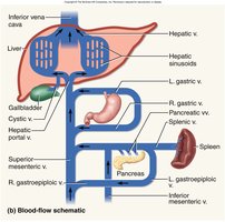
The liver receives blood from two sources: the hepatic artery (oxygen-rich) and the hepatic portal vein (nutrient-rich from the GI tract). Blood is filtered and then drained via the hepatic veins.
Proper hepatic artery: Supplies oxygenated blood.
Hepatic portal vein: Brings nutrient-rich blood from intestines.
Hepatic vein: Drains filtered blood to the inferior vena cava.
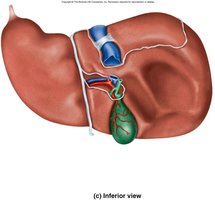
Inferior Surface of Liver
Key Structures
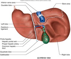
The inferior surface of the liver is closely associated with the gallbladder, hepatic portal vein, and proper hepatic artery.
Gallbladder: Adheres between right and quadrate lobes.
Inferior vena cava: Major venous drainage.
Hepatic portal vein and proper hepatic artery: Enter at porta hepatis.
Microscopic Anatomy of Liver
Hepatic Lobules and Cells
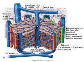
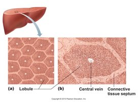
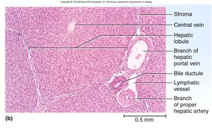
The liver is organized into hepatic lobules, each centered around a central vein and composed of hepatocytes separated by sinusoids.
Hepatocytes: Main functional cells, filter blood.
Sinusoids: Capillary channels for blood flow.
Kupffer cells: Hepatic macrophages that remove debris and pathogens.
Histology of Liver - Hepatic Triad
Portal Triad Components
Each corner of a hepatic lobule contains a portal triad, consisting of three structures:
Branch of hepatic portal vein
Branch of proper hepatic artery
Bile duct: Collects bile from canaliculi
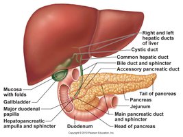
Ducts of Gallbladder, Liver, and Pancreas
Bile Flow and Regulation
Bile produced by the liver flows through a series of ducts before reaching the duodenum, regulated by the hepatopancreatic sphincter.
Bile canaliculi → bile ducts → right and left hepatic ducts → common hepatic duct → cystic duct → common bile duct → duodenum
Hepatopancreatic sphincter: Controls entry of bile and pancreatic juice into duodenum.
Gallbladder and Bile
Storage and Function
The gallbladder stores and concentrates bile, which is essential for fat digestion.
Bile: Yellow-green fluid containing bile pigments, bile salts, cholesterol, and phospholipids.
Bilirubin: Pigment from hemoglobin breakdown; converted to stercobilin by intestinal bacteria (gives feces brown color).
Gallbladder disease: May cause gray-white, fatty streaked stool.
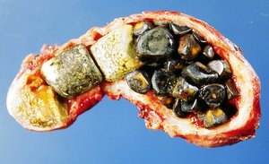
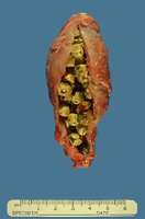
Gallstones
Formation and Clinical Features
Gallstones (biliary calculi) form when cholesterol crystallizes due to imbalance between cholesterol and bile salts, potentially blocking bile flow and causing pain.
Risk factors: Female, forty, fertile, fat (high cholesterol diet).
Treatment: Laparoscopic surgery or lithotripsy.
Gross Anatomy of Pancreas
Structure and Function
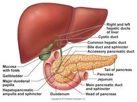
The pancreas is a retroperitoneal gland located posterior to the stomach, with both endocrine and exocrine functions.
Head, body, and tail: Anatomical regions.
Endocrine (islets): Secrete insulin and glucagon.
Exocrine (acini): Secrete pancreatic juice into duodenum.
Pancreatic duct: Opens at hepatopancreatic sphincter.
Pancreas - Acinar Cells
Secretory Function
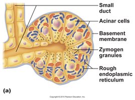
Acinar cells in the pancreas produce digestive enzymes stored in zymogen granules, and sodium bicarbonate to neutralize stomach acid.
Zymogen granules: Contain proteases, amylases, lipases, nucleases.
Sodium bicarbonate: Buffers acidic chyme.
Hormonal Control of Secretion
Regulation by CCK and Secretin
Digestive secretions are regulated by hormones released from the duodenum.
Cholecystokinin (CCK): Stimulates gallbladder contraction, pancreatic enzyme secretion, and relaxation of hepatopancreatic sphincter.
Secretin: Stimulates secretion of pancreatic juice, bicarbonate, and bile.
Small Intestine
Structure and Function
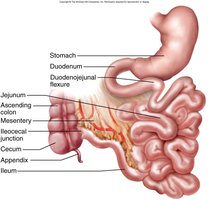
The small intestine is the primary site for chemical digestion and nutrient absorption, consisting of three segments: duodenum, jejunum, and ileum.
Duodenum: Neutralizes stomach acids, emulsifies fats, inactivates pepsin, receives pancreatic enzymes.
Jejunum: Most digestion and absorption occur here.
Ileum: Contains Peyer's patches (lymphatic nodules), ends at large intestine.
Small Intestine - Surface Area
Adaptations for Absorption
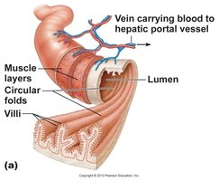
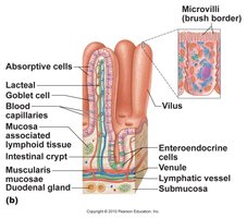
The small intestine has structural adaptations to maximize surface area for absorption.
Circular folds: Increase surface area.
Villi: Fingerlike projections containing blood vessels and lymphatics (lacteals).
Microvilli: Brush border with digestive enzymes.
Brunner’s glands: Secrete bicarbonate-rich mucus.
Peyer’s patches: Aggregated lymphoid follicles in ileum.
Intestinal Motility
Mixing and Propulsion
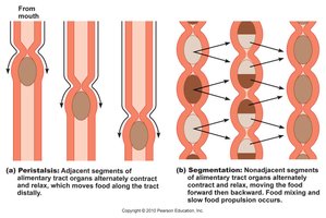
Intestinal motility involves peristalsis and segmentation to mix chyme and move it toward the large intestine.
Peristalsis: Wave-like contractions push chyme along.
Segmentation: Ring-like constrictions mix and churn contents.
Anatomy of Large Intestine
Segments and Features
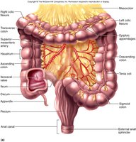
The large intestine absorbs water and electrolytes, forming feces. It consists of several segments and unique features.
Cecum, appendix, ascending, transverse, descending, sigmoid colon, rectum, anal canal
Ileocecal valve: Controls entry from ileum.
Haustrum: Pouch-like segments.
Tenia coli: Bands of longitudinal muscle.
Microscopic Anatomy of Large Intestine
Histological Features
The mucosa of the large intestine is simple columnar epithelium, except for the anal canal, which is stratified squamous. There are no villi or circular folds.
Intestinal crypts: Produce mucus only.
Teniae coli: Cause haustra formation.
Bacterial Flora
Role in Digestion
The large intestine is populated by diverse bacterial species that ferment undigested carbohydrates and synthesize vitamins.
Fermentation: Produces absorbable sugars.
Vitamin synthesis: B and K vitamins.
Flatus: Gas produced by bacteria and swallowed air.
Absorption and Motility in Large Intestine
Transit and Feces Formation
Transit time in the large intestine is 12-24 hours, during which water and electrolytes are reabsorbed and feces are formed.
Haustral contractions: Occur every 30 minutes.
Feces: Composed of water, bacteria, mucus, undigested fiber, fat, and sloughed cells.
Water Balance
Diarrhea and Constipation
Water absorption in the large intestine is critical for maintaining stool consistency.
Diarrhea: Too little water absorbed; rapid transit.
Constipation: Too much water absorbed; slow transit.
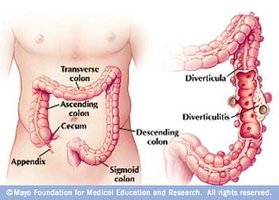
Diverticulitis
Pathology and Risk Factors
Diverticulitis is inflammation of diverticula in the colon, often associated with a diet high in fat and low in fiber.
Colon Cancer
Risk Factors and Detection
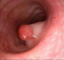
Colon cancer risk increases with polyps, low fiber/high fat diet, and genetic predisposition. Early detection via colonoscopy is crucial.
Polyps: Precancerous growths.
Colonoscopy: Recommended at age 50 or earlier with family history.
Lactose Intolerance
Causes and Management
Lactose intolerance is the inability to digest lactose due to declining lactase enzyme after age 4. Prevalence varies by ethnicity.
Symptoms: Gas, diarrhea, abdominal discomfort.
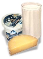
Management: Avoid lactose, consume yogurt/cheese, or take lactase supplements.
Breakdown Products of Digestion
Absorbed Nutrients
Digestion results in the breakdown of macromolecules into absorbable units:
Carbohydrates: Monosaccharides
Fats: Glycerol and fatty acids
Proteins: Amino acids
DNA/RNA: Nucleotides
Vitamins: Absorbed unchanged
Carbohydrate Digestion
Enzymes and Absorption
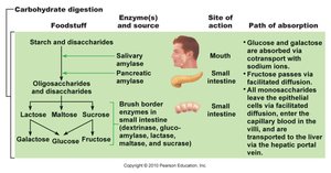
Carbohydrate digestion begins in the mouth and is completed in the small intestine.
Salivary amylase: Denatured in stomach (pH < 4.5).
Pancreatic amylase: Completes starch digestion in small intestine.
Brush border enzymes: Final breakdown to monosaccharides.
Foodstuff | Enzyme(s) and Source | Site of Action | Path of Absorption |
|---|---|---|---|
Starch and disaccharides | Salivary amylase, pancreatic amylase | Mouth, small intestine | Glucose and galactose absorbed via cotransport with sodium ions; fructose via facilitated diffusion |
Lactose, maltose, sucrose | Brush border enzymes (dextrinase, glucoamylase, lactase, maltase, sucrase) | Small intestine | Monosaccharides enter capillary blood in villi, transported to liver via hepatic portal vein |
Protein Digestion
Enzymes and Absorption
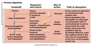
Protein digestion starts in the stomach and continues in the small intestine.
Pepsin: Active in stomach (pH 1.5-3.5), inactivated in duodenum.
Trypsin and chymotrypsin: Digest proteins in small intestine.
Foodstuff | Enzyme(s) and Source | Site of Action | Path of Absorption |
|---|---|---|---|
Protein | Pepsin (stomach glands) | Stomach | Amino acids absorbed by cotransport with sodium ions |
Large polypeptides | Pancreatic enzymes (trypsin, chymotrypsin, carboxypeptidase) | Small intestine | Amino acids enter capillary blood in villi, transported to liver via hepatic portal vein |
Small polypeptides, small peptides | Brush border enzymes (aminopeptidase, carboxypeptidase, dipeptidase) | Small intestine | Same as above |
Fat Digestion
Absorption Pathways
Most fats are transported to the blood via lymphatic lacteals, while some enter capillary blood and are transported to the liver.
Nucleic Acid Digestion
Breakdown and Absorption
Nucleic acids are digested into nucleotides, which are absorbed in the small intestine.
Summary Table: Digestive Enzymes and Absorption
Key Digestive Processes
Macromolecule | Enzymes | Site of Action | Absorbed Form |
|---|---|---|---|
Carbohydrates | Amylases, brush border enzymes | Mouth, small intestine | Monosaccharides |
Proteins | Pepsin, trypsin, chymotrypsin, peptidases | Stomach, small intestine | Amino acids |
Fats | Lipases | Small intestine | Fatty acids, glycerol |
Nucleic acids | Nucleases | Small intestine | Nucleotides |
Key Equations
Relevant Biochemical Reactions
Carbohydrate hydrolysis:
Protein hydrolysis:
Fat hydrolysis:
Additional info: Some details were expanded for academic completeness, such as the summary tables and biochemical equations.
