 Indietro
Digestive System: Mechanisms and Processes of Digestion and Absorption
Guida di studio - Note intelligenti
 Appunti personalizzati basati sui tuoi materiali, ampliati con definizioni chiave, esempi e contesto.
Mechanism of HCl Formation in the Stomach
Parietal Cell Function and Acid Secretion
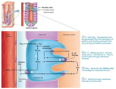
The parietal cells of the gastric glands are responsible for secreting hydrochloric acid (HCl) into the stomach lumen, a process essential for digestion and defense against pathogens.
Carbonic Acid Breakdown: Carbon dioxide (CO2) combines with water (H2O) in parietal cells, catalyzed by carbonic anhydrase, forming carbonic acid (H2CO3), which dissociates into H+ and HCO3-.
Proton Pump: H+ ions are pumped into the stomach lumen via the H+/K+ ATPase (proton pump).
Alkaline Tide: HCO3- is exported to the blood via a Cl-/HCO3- antiporter, causing a temporary increase in blood alkalinity (alkaline tide).
Formation of HCl: Cl- ions are pumped into the lumen, where they combine with H+ to form HCl.
Gastric Contractile Activity
Peristalsis and Chyme Movement
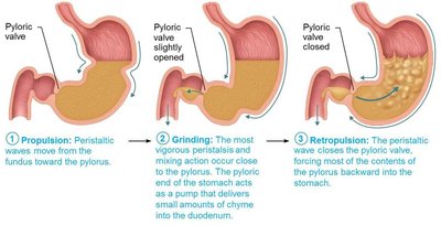
The stomach uses rhythmic contractions to mix and propel its contents toward the duodenum, facilitating digestion and regulating gastric emptying.
Peristaltic Waves: Move toward the pylorus at a rate of three per minute, driven by the basic electrical rhythm (BER) set by enteric pacemaker cells.
Pacemaker Cells: Linked by gap junctions, ensuring coordinated contraction of the muscularis.
Distension and Gastrin: Increase the force of contraction.
Chyme Delivery: About 3 ml of chyme is delivered to the duodenum per contraction; the rest is forced backward when the pyloric valve closes.
Selective Passage: Only liquids and small particles pass through the pyloric valve.
Regulation of Gastric Emptying
Duodenal Control and Feedback Mechanisms
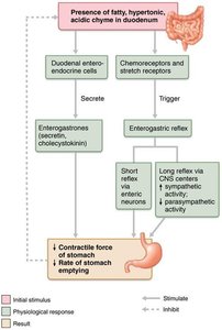
The duodenum regulates the rate at which chyme enters from the stomach, preventing overfilling and ensuring optimal digestion and absorption.
Duodenal Receptors: Respond to stretch and chemical signals.
Enterogastric Reflex: Inhibits gastric secretion and slows duodenal filling.
Enterogastrones: Hormones (e.g., secretin, cholecystokinin) that inhibit gastric activity.
Emptying Time: Stomach empties in about 4 hours; fatty chyme increases this time.
Carbohydrate-rich Chyme: Moves quickly through the duodenum.
Liver, Gallbladder, and Pancreas
Accessory Organs of Digestion
The liver, gallbladder, and pancreas are essential accessory organs that support digestion in the small intestine.
Liver: Produces bile, a fat emulsifier.
Gallbladder: Stores and concentrates bile.
Pancreas: Produces digestive enzymes and bicarbonate to neutralize stomach acid.
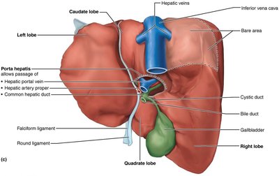
Gross Anatomy of the Liver
Lesser Omentum: Anchors liver to stomach.
Porta Hepatis: Entry point for hepatic artery and vein.
Bile Ducts: Common hepatic duct leaves liver, cystic duct connects to gallbladder, and bile duct is formed by their union.
Microscopic Anatomy of the Liver
Hepatocytes: Highly metabolic cells with abundant ER, Golgi, peroxisomes, and mitochondria.
Functions: Produce bile, process nutrients, store vitamins, detoxify blood, and synthesize plasma proteins.
Stellate Macrophages: Remove debris and old RBCs from blood.
Bile Composition and Enterohepatic Circulation
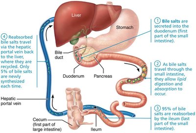
Bile: Yellow-green, alkaline solution containing bile salts, bilirubin, cholesterol, triglycerides, phospholipids, and electrolytes.
Bile Salts: Cholesterol derivatives aiding fat emulsification and absorption.
Bilirubin: Waste product from RBC breakdown; converted to stercobilin in intestine, giving feces its brown color.
Enterohepatic Circulation: Bile salts are reabsorbed in the ileum and recycled to the liver; only 5% are newly synthesized each cycle.
Gallstones and Gallbladder Function
Gallstone Formation and Effects
Gallstones: Formed by excess cholesterol or insufficient bile salts; can obstruct bile flow and cause pain.
Obstructive Jaundice: Blockage leads to accumulation of bile pigments in blood, resulting in jaundice.
Treatment: Includes drugs, lithotripsy, laser vaporization, or surgery.
Pancreas: Structure and Function
Exocrine and Endocrine Roles
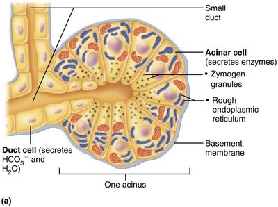
Exocrine Function: Acini produce digestive enzymes; duct cells secrete water and bicarbonate.
Endocrine Function: Islet cells secrete insulin and glucagon.
Composition of Pancreatic Juice
Volume: 1200–1500 ml/day, alkaline (pH 8).
Components: Water, electrolytes (mainly HCO3-), digestive enzymes (proteases, amylase, lipases, nucleases).
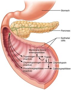
Protease Activation: Proteases are secreted in inactive forms and activated in the duodenum by enteropeptidase.
Regulation of Bile and Pancreatic Secretions
Neural and Hormonal Controls
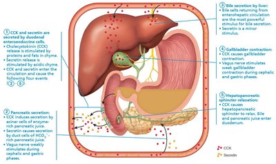
Hormones: Cholecystokinin (CCK) and secretin stimulate secretion.
Bile Secretion: Increased by enterohepatic circulation and secretin.
Pancreatic Secretion: Stimulated by CCK and secretin in response to acidic and fatty chyme.
Small Intestine: Structure and Function
Major Organ of Digestion and Absorption
Length: 2–4 m, subdivided into duodenum, jejunum, and ileum.
Blood Supply: Superior mesenteric artery and hepatic portal vein.
Nerve Supply: Parasympathetic (vagus nerve) and sympathetic (thoracic splanchnic nerves).
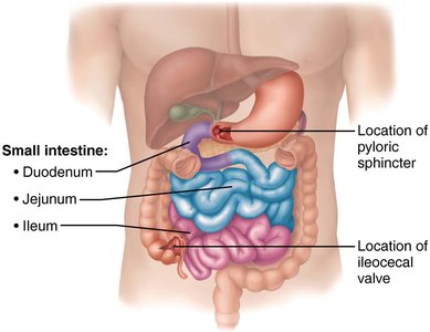
Modifications for Absorption
Surface Area: Increased 600× to ~200 m2 by structural modifications.
Circular Folds: Permanent folds that slow chyme movement for absorption.
Villi: Fingerlike projections containing capillaries and lacteals for nutrient absorption.
Microvilli: Cytoplasmic extensions forming the brush border with digestive enzymes.
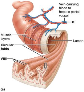
Histology of Small Intestine Wall
Intestinal Crypts: Tubular glands between villi.
Cell Types:
Enterocytes: Absorptive cells with microvilli; absorb nutrients and electrolytes.
Goblet Cells: Secrete mucus.
Enteroendocrine Cells: Secrete hormones (CCK, secretin).
Paneth Cells: Secrete antimicrobial agents.
Stem Cells: Continuously divide to renew epithelium.
MALT: Mucosa-associated lymphoid tissue protects against microorganisms; includes Peyer's patches and plasma cells secreting IgA.
Submucosa: Areolar tissue with duodenal glands secreting alkaline mucus.
Intestinal Juice
Volume: 1–2 L/day, slightly alkaline and isotonic with blood plasma.
Composition: Water and mucus, secreted by duodenal glands and goblet cells.
Motility of the Small Intestine
Segmentation and Peristalsis
Segmentation: Most common after a meal; mixes and moves contents toward ileocecal valve.
Peristalsis: Occurs between meals, moving remnants toward large intestine.
Regulation: Parasympathetic increases motility; sympathetic decreases it.
Large Intestine: Structure and Function
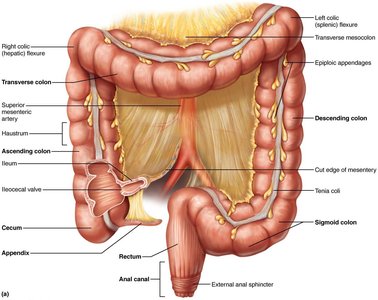
Subdivisions and Anatomy
Cecum: First part; receives chyme from ileum.
Appendix: Contains lymphoid tissue; can store bacteria and is susceptible to blockages.
Colon: Ascending, transverse, descending, and sigmoid regions.
Appendicitis
Definition: Acute inflammation due to blockage and infection.
Symptoms: Pain, loss of appetite, nausea, vomiting.
Treatment: Surgical removal or antibiotics.
Bacterial Flora
Composition: 1000+ types of bacteria, outnumbering human cells.
Functions: Fermentation of indigestible carbohydrates, vitamin synthesis (B and K), suppression of pathogens.
Health Impact: Gut bacteria influence body weight, disease susceptibility, and mood.
Homeostatic Imbalance
Antibiotic-associated Diarrhea: Caused by Clostridium difficile; may require fecal transplants.
Digestive Processes in the Large Intestine
Residue: Remains for 12–24 hours; no food breakdown except by bacteria.
Absorption: Vitamins, water, and electrolytes reclaimed.
Primary Functions: Absorb water, eliminate feces, harvest vitamins.
Defecation
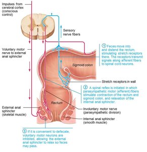
Mass Movements: Force feces toward rectum.
Defecation Reflex: Initiated by distension; parasympathetic signals stimulate contraction and relaxation of sphincters.
Conscious Control: Allows relaxation of external anal sphincter.
Mechanism of Digestion and Absorption
Overview of Digestion
Digestion: Catabolic process breaking macromolecules into monomers for absorption.
Enzymes: Carry out hydrolysis reactions.
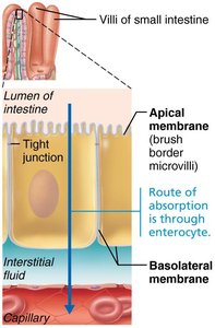
Absorption: Movement of substances from gut lumen into body; most nutrients absorbed before chyme reaches ileum.
Tight Junctions: Ensure molecules pass through epithelial cells.
Digestion and Absorption of Carbohydrates
Enzymatic Breakdown and Transport
Only Monosaccharides: Can be absorbed.
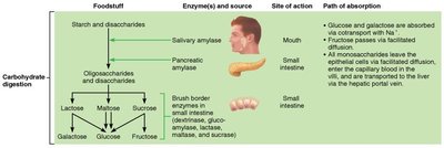
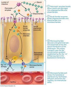
Starch and Disaccharides: Broken down by salivary and pancreatic amylase, then brush border enzymes.
Final Products: Glucose, fructose, galactose.
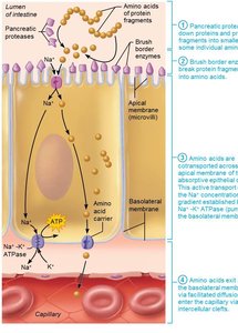
Transport: Monosaccharides are cotransported with Na+ across apical membrane and exit via facilitated diffusion.
Digestion and Absorption of Proteins
Enzymatic Breakdown and Transport
Sources: Dietary proteins, digestive enzymes, and mucosal cell proteins.
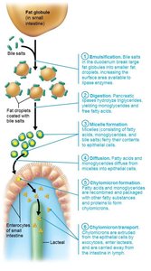
Digestion: Begins in stomach with pepsin; continues in small intestine with pancreatic proteases and brush border enzymes.
Final Products: Amino acids, dipeptides, tripeptides.
Transport: Amino acids are cotransported with Na+ or H+ and exit via facilitated diffusion.
Digestion and Absorption of Lipids
Emulsification, Digestion, and Transport
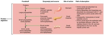
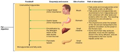
Emulsification: Bile salts break large fat globules into smaller ones.
Digestion: Pancreatic lipases break down fats into monoglycerides and free fatty acids.
Micelle Formation: Digestion products are coated with bile salts and lecithin.
Diffusion: Lipid products cross epithelial membrane.
Chylomicron Formation: Lipid products are packaged and transported via lymphatic lacteals.
Short-chain Fatty Acids: Diffuse directly into blood.
Digestion and Absorption of Nucleic Acids
Enzymatic Breakdown and Transport
Digestion: Pancreatic nucleases hydrolyze DNA and RNA to nucleotide monomers.
Brush Border Enzymes: Nucleosidases and phosphatases break nucleotides into nitrogenous bases, pentose sugars, and phosphate ions.
Transport: Breakdown products are actively transported by special carriers.
Vitamin, Electrolyte, and Water Absorption
Absorption Mechanisms
Fat-soluble Vitamins: (A, D, E, K) carried by micelles and diffuse into cells.
Water-soluble Vitamins: (C, B) absorbed by diffusion or transporters; B12 requires intrinsic factor.
Electrolytes: Most ions transported actively; iron and calcium absorbed in duodenum, regulated by need and hormones.
Water: 95% absorbed in small intestine by osmosis; uptake coupled with solute absorption.
Homeostatic Imbalance: Malabsorption
Causes: Interference with bile or pancreatic juice delivery, damaged mucosa, celiac disease.
Celiac Disease: Immune reaction to gluten damages villi and brush border; treated by eliminating gluten.
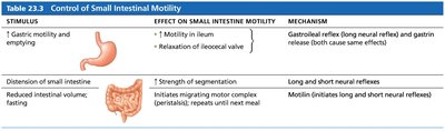
Summary Table: Control of Small Intestinal Motility
Stimulus, Effect, and Mechanism
Stimulus | Effect on Small Intestine Motility | Mechanism |
|---|---|---|
Gastric motility and emptying | ↑ Motility in ileum; relaxation of ileocecal valve | Gastroileal reflex (long neural reflex) and gastrin release |
Distension of small intestine | ↑ Strength of segmentation | Long and short neural reflexes |
Reduced intestinal volume; fasting | Initiates migrating motor complex (peristalsis) | Motilin (initiates long and short neural reflexes) |
Summary Table: Digestion and Absorption of Carbohydrates, Proteins, and Lipids
Carbohydrate Digestion
Foodstuff | Enzyme(s) and Source | Site of Action | Path of Absorption |
|---|---|---|---|
Starch and disaccharides | Salivary amylase, pancreatic amylase | Mouth, small intestine | Glucose and galactose absorbed via Na+ cotransport; fructose via facilitated diffusion |
Oligosaccharides and disaccharides | Brush border enzymes (dextrinase, glucoamylase, lactase, maltase, sucrase) | Small intestine | Monosaccharides leave epithelial cells via facilitated diffusion |
Protein Digestion
Foodstuff | Enzyme(s) and Source | Site of Action | Path of Absorption |
|---|---|---|---|
Proteins | Pepsin (stomach glands), pancreatic enzymes (trypsin, chymotrypsin, carboxypeptidase), brush border enzymes | Stomach, small intestine | Amino acids absorbed via Na+ cotransport; dipeptides and tripeptides hydrolyzed within cells |
Fat Digestion
Foodstuff | Enzyme(s) and Source | Site of Action | Path of Absorption |
|---|---|---|---|
Unemulsified triglycerides | Lingual lipase, gastric lipase, bile salts, pancreatic lipases | Mouth, stomach, small intestine | Fatty acids and monoglycerides enter intestinal cells via diffusion; chylomicrons transported via lymph |
