 Indietro
Digestive System Part II: Structure, Function, and Histology
Guida di studio - Note intelligenti
 Appunti personalizzati basati sui tuoi materiali, ampliati con definizioni chiave, esempi e contesto.
Digestive System Overview
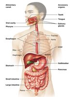
Alimentary Canal and Accessory Organs
The digestive system consists of the alimentary canal and accessory organs, each playing a vital role in digestion and absorption.
Alimentary canal: Includes the esophagus, stomach, small intestine, and large intestine.
Accessory organs: Liver, gallbladder, and pancreas, which produce and store substances essential for digestion.
Large Intestine Structure
Major Divisions of the Large Intestine
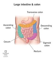
The large intestine is divided into four main regions, each with distinct anatomical and functional characteristics.
Cecum: The initial pouch-like segment.
Colon: Subdivided into ascending, transverse, descending, and sigmoid sections.
Rectum: Terminal straight portion.
Anal Canal: Final segment leading to the anus.
Taeniae coli: Longitudinal ribbons of smooth muscle that contract to form haustra.
Haustra: Segmented pouches that give the large intestine its characteristic appearance.
Pancreas and Biliary System
Pancreatic Ducts and Bile Flow
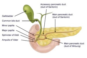
The pancreas and biliary system work together to deliver digestive enzymes and bile to the duodenum.
Main pancreatic duct: Transports digestive enzymes to the duodenum via the major duodenal papilla.
Common bile duct: Formed by the union of the common hepatic duct and cystic duct, it carries bile to the duodenum.
Major duodenal papilla: Entry point for both bile and pancreatic juices into the small intestine.
Liver and Gallbladder Functions
The liver and gallbladder are essential for bile production, storage, and release.
Liver: Produces bile, which contains bilirubin (a waste product) and bile salts for fat emulsification.
Gallbladder: Stores and concentrates bile, releasing it via the cystic duct when needed.
Hepatic portal system: Transports nutrient-rich blood from the digestive tract to the liver for processing.
Biliary Duct Anatomy
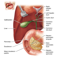
The ducts of the liver and gallbladder form a complex network for bile transport.
Right and left hepatic ducts: Merge to form the common hepatic duct.
Common hepatic duct: Transports bile from the liver.
Cystic duct: Connects the gallbladder to the common bile duct.
Common bile duct: Formed by the union of the common hepatic and cystic ducts; joins with the main pancreatic duct.
Histology of the Large Intestine
Microscopic Features
The large intestine is characterized by its abundance of goblet cells and specific epithelial structures.
Goblet cells: Specialized for mucus secretion, aiding in the passage of feces.
Simple columnar epithelium: Lines the mucosa, facilitating absorption and secretion.
Muscularis mucosae and submucosa: Support the mucosal layer and provide structural integrity.
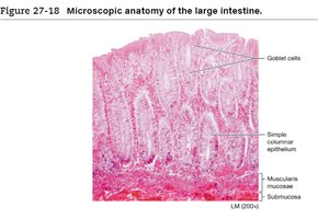
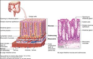
Goblet Cell Abundance
Goblet cells are a key identifier in large intestine histology, appearing in high numbers throughout the mucosa. 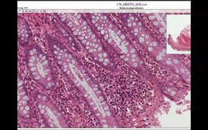
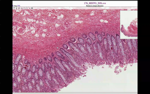
Liver Histology
Liver Lobule Structure
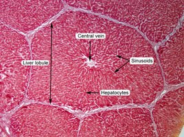
The liver is organized into lobules, each with a central vein and portal triads at the corners.
Central vein: Located at the center of each lobule, drains blood from the liver.
Portal triad: Consists of a bile duct (simple columnar epithelium), portal arteriole (muscular layer), and portal venule (large, thin-walled).
Sinusoids: Capillary-like spaces between hepatocytes, filtering blood.
Liver Blood Flow and Bile Secretion
Blood and Bile Pathways
Bile secretion: Hepatocytes produce bile, which travels through bile canaliculi to bile ducts.
Blood flow: Blood enters via the portal triad, passes through sinusoids, drains into the central vein, then hepatic vein, and finally the inferior vena cava.
Pancreas Histology
Microscopic Structure
The pancreas contains acinar cells and ducts specialized for enzyme production and transport.
Acinar cell: Individual cell responsible for enzyme secretion.
Acinus (pl. acini): Cluster of acinar cells surrounding a duct.
Pancreatic ducts: Lined with cuboidal epithelial cells, transport enzymes to the duodenum.
Comparative Histology: Stomach, Small Intestine, and Large Intestine
Key Differences
Stomach: Contains gastric glands and a thick muscularis layer.
Small intestine: Features villi and microvilli for increased absorption.
Large intestine: Abundant goblet cells, no villi, and a smooth mucosal surface.
Summary Table: Digestive System Structures and Functions
Structure | Main Function | Key Histological Feature |
|---|---|---|
Stomach | Mechanical and chemical digestion | Gastric glands, thick muscularis |
Small Intestine | Absorption of nutrients | Villi, microvilli |
Large Intestine | Water absorption, feces formation | Goblet cells, haustra |
Liver | Bile production, metabolism | Lobules, portal triad, central vein |
Pancreas | Enzyme secretion | Acinar cells, ducts |
Gallbladder | Bile storage | Simple columnar epithelium |
Additional info: Academic context was added to clarify histological features, duct anatomy, and comparative structure-function relationships.
