 Indietro
Digestive System: Structure, Function, and Nutrient Absorption
Guida di studio - Note intelligenti
 Appunti personalizzati basati sui tuoi materiali, ampliati con definizioni chiave, esempi e contesto.
Overview of Digestive System Functions
Major Functions of the Digestive System
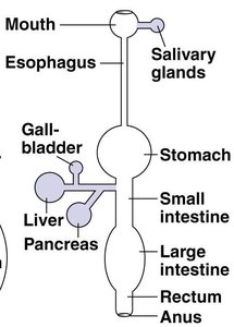
The digestive system is responsible for breaking down food, absorbing nutrients, and eliminating waste. Its functions include ingestion, secretion, propulsion, digestion (mechanical and chemical), absorption, and defecation.
Ingestion: Intake of food into the mouth.
Secretion: Release of digestive fluids and enzymes.
Propulsion: Movement of food through the tract, including peristalsis.
Digestion:
Mechanical: Physical breakdown (mastication, churning, segmentation).
Chemical: Enzymatic hydrolysis of macromolecules.
Absorption: Uptake of nutrients into blood or lymph.
Defecation: Elimination of indigestible substances.
Small Intestine: Structure and Function
Anatomy of the Small Intestine
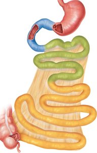
The small intestine is the primary site for chemical digestion and nutrient absorption. It extends from the pyloric sphincter to the ileocecal sphincter and is divided into three regions: duodenum, jejunum, and ileum.
Duodenum: Receives chyme from the stomach and secretions from the liver, gallbladder, and pancreas.
Jejunum: Major site for nutrient absorption.
Ileum: Absorbs remaining nutrients and vitamin B12; contains Peyer's patches (MALT).
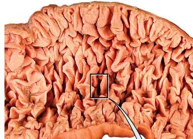
Circular folds: Permanent ridges in mucosa and submucosa that increase surface area for absorption.
Histology of the Small Intestine
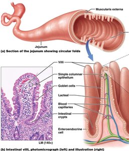
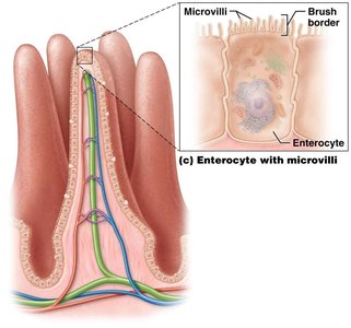
The mucosal layer is folded into villi, each containing a lacteal and blood supply. The submucosal layer contains duodenal glands and MALT.
Villi: Finger-like projections that increase surface area.
Microvilli: Apical surface of enterocytes forms a brush border, further increasing surface area and containing digestive enzymes.
Goblet cells: Secrete mucus for lubrication.
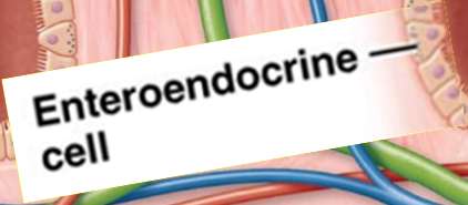
Enteroendocrine cells: Secrete hormones (secretin, CCK, GIP) to regulate digestive activity.
Motility in the Small Intestine
Motility is essential for mixing and propelling contents.
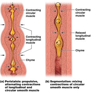
Segmentation: Mixing contractions of circular smooth muscle (intestinal churning).
Peristalsis: Propulsive, alternating contractions of longitudinal and circular smooth muscle.
Large Intestine: Structure and Function
Anatomy and Functions of the Large Intestine
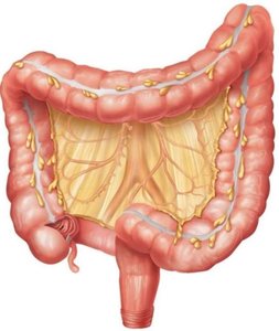
The large intestine completes water absorption, produces certain vitamins, forms and expels feces. It extends from the ileocecal sphincter to the anus and is divided into cecum, colon (ascending, transverse, descending, sigmoid), rectum, and anal canal.
Appendix: Inferior to cecum.
Colon: Four regions; haustra are pouches formed by taeniae coli.
Histology of the Large Intestine
Mucosa: Increased goblet cells, no villi or circular folds.
Absorptive cells: Colonocytes absorb water and electrolytes.
Muscularis: Longitudinal muscle forms taeniae coli, creating haustra.
Defecation Reflex
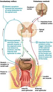
The defecation reflex is triggered by stretch receptors in the sigmoid colon. Voluntary contractions of the diaphragm and abdominal muscles aid elimination, and relaxation of the external anal sphincter allows defecation.
Feces: Mixture of undigested food, bacteria, salts, water, and epithelial cells.
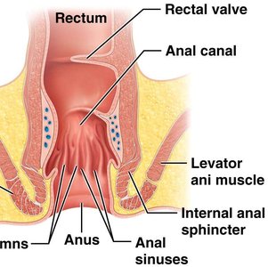
Internal and external anal sphincters: Control passage of feces.
Accessory Organs: Pancreas, Liver, and Gallbladder
Pancreas: Structure and Digestive Functions
The pancreas is located posterior to the stomach and has both endocrine (islets) and exocrine (acini) functions.
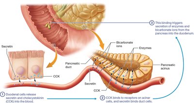
Exocrine function: Secretes digestive enzymes and bicarbonate ions into the duodenum.
Enzymes: Pancreatic amylase (starch), trypsin/chymotrypsin/carboxypeptidase (proteins), pancreatic lipase (fats), ribonuclease/deoxyribonuclease (nucleic acids).
Liver and Gallbladder: Structure and Functions
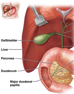
The liver is the second largest organ, divided into left and right lobes. Its only digestive function is bile production, which emulsifies fats. The gallbladder stores and releases bile.
Liver: Processes nutrients, detoxifies substances, stores glycogen.
Gallbladder: Receives, stores, and releases excess bile.
Bile ducts: Transport bile from liver and gallbladder to duodenum.
Liver Histology and Blood Supply
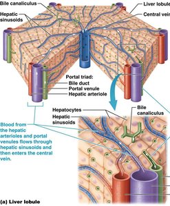
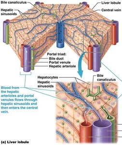
Hepatocytes: Liver cells arranged in lobules around a central vein.
Portal triad: Contains bile duct, hepatic arteriole (O2-rich blood), and hepatic portal venule (nutrient-rich, O2-poor blood).
Blood flow: All blood drains into hepatic sinusoids and then to the central vein.
Nutrient Digestion and Absorption
Enzymatic Hydrolysis and Digestive Enzymes
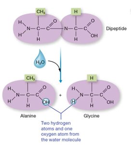
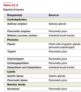
Enzymatic hydrolysis breaks down macromolecules into absorbable units. Digestive enzymes are specific to substrates and are activated in different regions of the GI tract.
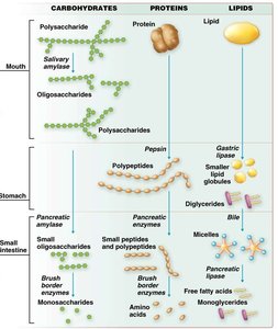
Carbohydrates: Digested by salivary amylase, pancreatic amylase, and brush border enzymes.
Proteins: Digested by pepsin, trypsin, chymotrypsin, carboxypeptidase, and brush border enzymes.
Lipids: Digested by gastric and pancreatic lipase, aided by bile.
Nucleic acids: Digested by pancreatic nucleases.
Enzyme(s) | Source |
|---|---|
Carbohydrates | |
Salivary amylase | Salivary glands |
Pancreatic amylase | Pancreatic juice |
Maltase, sucrase, lactase | Intestinal brush border |
Proteins | |
Pepsin | Chief cells of gastric glands |
Trypsin | Pancreatic juice |
Chymotrypsin | Pancreatic juice |
Carboxypeptidase | Pancreatic juice |
Dipeptidase, tripeptidase | Intestinal brush border |
Lipids | |
Gastric lipase | Gastric glands |
Pancreatic lipase | Pancreatic juice |
Nucleic Acids | |
Nucleases | Pancreatic juice |
Digestion and Absorption of Nutrients
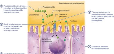
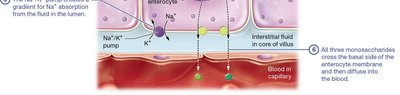
Carbohydrates: Digested into monosaccharides (glucose, galactose, fructose) and absorbed into blood capillaries.
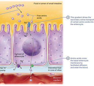
Proteins: Digested into amino acids and absorbed into blood capillaries.
Lipids: Digested into fatty acids and monoglycerides, absorbed into lacteals as chylomicrons.
Nucleic acids, water, electrolytes, vitamins: Absorbed into blood capillaries.
Absorption of Water, Electrolytes, and Vitamins
The intestines absorb large quantities of water, electrolytes, and vitamins.
Water: Most is absorbed in the small intestine; remainder in the large intestine.
Electrolytes: Sodium, potassium, calcium ions absorbed in the small intestine.
Vitamins:
Fat-soluble (A, D, E, K): Packaged into micelles and absorbed with lipids.
Water-soluble (B, C): Mostly absorbed in the small intestine.
Vitamin B12: Must bind to intrinsic factor for absorption in the ileum.
Example: More than 9 liters of water enter the small intestine daily, with about 8 liters absorbed by enterocytes and only 0.1 liters excreted in feces. Additional info: The absorption of nutrients is highly regulated and depends on the presence of specific transporters and enzymes in the intestinal epithelium. ----------------------------------------
