 Indietro
Digestive System: Structure, Function, and Processes
Guida di studio - Note intelligenti
 Appunti personalizzati basati sui tuoi materiali, ampliati con definizioni chiave, esempi e contesto.
Overview of the Digestive System
General Structure and Organization
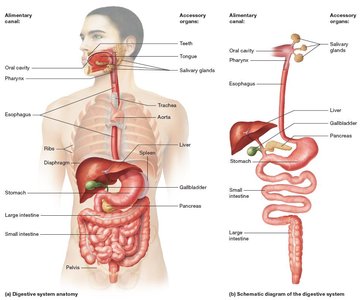
The digestive system is a complex set of organs responsible for breaking down food into nutrients that can be absorbed and utilized by the body. It consists of the alimentary canal (gastrointestinal tract) and accessory organs. The alimentary canal is a continuous tube extending from the oral cavity to the anus, while accessory organs assist in digestion but are not part of the canal itself.
Alimentary canal: Oral cavity, pharynx, esophagus, stomach, small intestine, large intestine
Accessory organs: Teeth, tongue, salivary glands, liver, gallbladder, pancreas
Food passes directly through the alimentary canal; accessory organs aid digestion (e.g., teeth and tongue physically manipulate food, glands secrete enzymes).
Basic Digestive Functions and Processes
The digestive system performs several essential functions to maintain homeostasis and provide nutrients:
Ingestion: Intake of food and water, primarily through the mouth.
Secretion: Release of digestive enzymes, hormones, and mucus by both endocrine and exocrine glands.
Propulsion: Movement of food and water through the canal, mainly via peristalsis (rhythmic contractions of smooth muscle).
Digestion:
Mechanical digestion: Physical breakdown of food (chewing, mixing).
Chemical digestion: Enzymatic breakdown of food into small molecules.
Absorption: Transfer of digested nutrients, water, electrolytes, and vitamins into blood or lymph.
Defecation: Elimination of indigestible materials and metabolic wastes as feces.
Organization of Abdominopelvic Digestive Organs
Peritoneal Membranes and Mesenteries
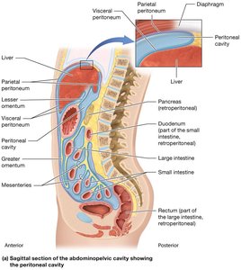
Digestive organs in the abdominopelvic cavity are surrounded by peritoneal membranes, the largest serous membrane in the body. These membranes provide support, reduce friction, and house blood vessels and nerves.
Parietal peritoneum: Lines the inner surface of the body wall.
Visceral peritoneum (serosa): Covers abdominal organs.
Peritoneal cavity: Space between the two layers, filled with serous fluid.
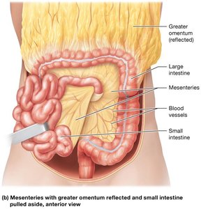
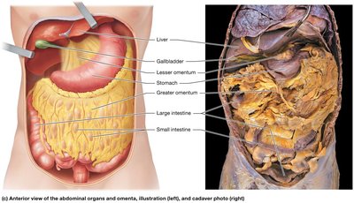
Mesenteries: Folds of visceral peritoneum that anchor organs, contain vessels and nerves, and maintain organ position.
Greater omentum: Four-layered fold extending from the stomach.
Lesser omentum: Connects stomach to liver.
Mesocolon: Anchors the large intestine.
Intraperitoneal organs: Completely within the peritoneal cavity.
Retroperitoneal organs: Partly or completely outside the peritoneal cavity.
Blood Supply and Innervation
Splanchnic circulation: Arterial supply from branches of the abdominal aorta (celiac trunk, superior and inferior mesenteric arteries).
Venous drainage via hepatic portal vein to the liver, then hepatic veins to the inferior vena cava.
Nervous supply from autonomic nervous system (sympathetic and parasympathetic) and enteric nervous system (ENS).
ENS plexuses: Submucosal (Meissner’s) plexus and Myenteric (Auerbach’s) plexus regulate secretion and motility.
Histology of the Alimentary Canal
General Tissue Layers
Most regions of the alimentary canal share a four-layered structure:
Mucosa: Inner epithelium, lamina propria (loose connective tissue), muscularis mucosae (smooth muscle).
Submucosa: Dense irregular connective tissue, blood and lymphatic vessels, glands, and submucosal plexus.
Muscularis externa: Two layers of smooth muscle (inner circular, outer longitudinal), regulated by myenteric plexus.
Serosa (visceral peritoneum): Simple squamous epithelium and loose connective tissue (in peritoneal cavity).
Adventitia: Dense irregular connective tissue (outside peritoneal cavity).
Regulation of Motility
Nervous and Endocrine Control
Motility: Movement of food via swallowing, churning, peristalsis, and defecation.
Autonomic nervous system:
Sympathetic division inhibits motility.
Parasympathetic division stimulates motility.
Enteric nervous system (ENS): Local control via short reflexes.
Endocrine system: Hormones and paracrines produced by digestive organs regulate motility and secretion.
The Oral Cavity, Pharynx, and Esophagus
Structure of the Oral Cavity
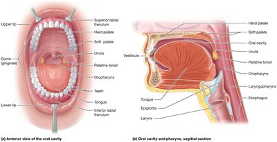
The oral cavity is the entry point for food and houses accessory organs such as teeth and tongue. It is bounded by the palate, cheeks, lips, and gums.
Cheeks: Buccinator muscles, lined by stratified squamous nonkeratinized epithelium.
Lips: Orbicularis oris muscle, covered with keratinized epithelium.
Gums (gingivae): Stratified squamous epithelium over connective tissue.
Palate:
Hard palate (anterior): Stratified squamous epithelium over bone.
Soft palate (posterior): Stratified squamous epithelium over skeletal muscle; includes uvula.
The Teeth and Mastication
Teeth: Located in alveoli of mandible and maxilla, held by periodontal ligament.
Mastication: Chewing increases surface area for enzymatic digestion.
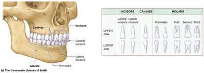
Types of teeth:
Incisors: Cutting
Canines: Tearing
Premolars and molars: Grinding
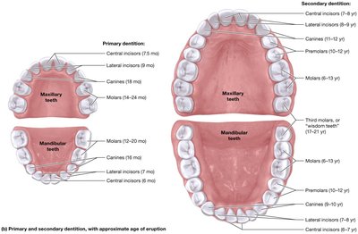
Primary dentition: 20 deciduous teeth (erupt by age 2).
Secondary dentition: 32 permanent teeth (erupt by age 12-21).
Tooth Structure
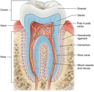
Crown: Enamel (hardest substance in body), covers dentin and pulp.
Root: Cementum covers dentin, anchors tooth.
Pulp cavity: Contains blood vessels and nerves.
Root canal: Extension of pulp cavity into root.
Dental Caries
Caries (cavities): Caused by bacterial metabolism of sugars, producing acids that dissolve enamel.
Prevention: Fluoride, brushing, flossing, sealants, reduced sugar intake.
The Tongue
Tongue: Skeletal muscle covered with stratified squamous epithelium.
Papillae: Filiform (texture), fungiform, circumvallate, foliate (taste buds).
Muscle groups:
Extrinsic: Position
Intrinsic: Shape and size
Salivary Glands
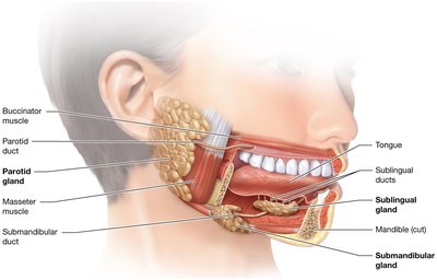
Three pairs: Parotid (serous), submandibular (mostly serous), sublingual (mostly mucous).
Acinar cells: Serous (enzymes), mucous (mucus).
Saliva: Water, electrolytes, mucus, salivary amylase (carbohydrate digestion), lysozyme (antibacterial), IgA (immune), bicarbonate (neutralizes acid).
Salivation: Controlled by parasympathetic and sympathetic nervous systems.
Pharynx and Esophagus
Pharynx: Oropharynx and laryngopharynx are part of alimentary canal; lined with stratified squamous epithelium.
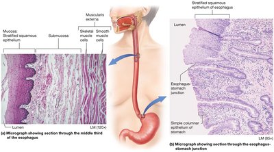
Esophagus: Muscular tube (25 cm), transports bolus to stomach; mucosa lined with stratified squamous epithelium.
Sphincters: Upper esophageal sphincter (entry), gastroesophageal sphincter (exit to stomach).
Motility: Peristalsis propels bolus; thick epithelium protects from abrasion.
Swallowing (Deglutition)
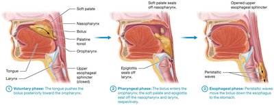
Three phases:
Voluntary: Tongue pushes bolus to oropharynx.
Pharyngeal: Soft palate and epiglottis seal off nasopharynx and larynx; involuntary contraction.
Esophageal: Peristaltic waves move bolus to stomach.
Control: Neural (cerebral cortex, medulla, ENS); ANS influences esophageal phase.
The Stomach
Gross Anatomy
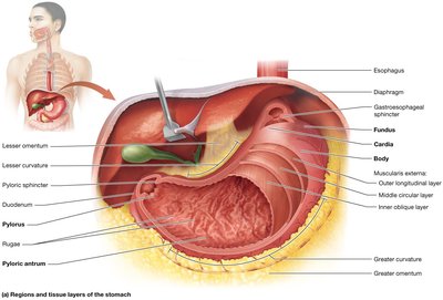
Regions: Cardia, fundus, body, pyloric antrum, pylorus.
Curvatures: Greater (left), lesser (right).
Rugae: Folds allowing expansion.
Pyloric sphincter: Controls flow to duodenum.
Histology
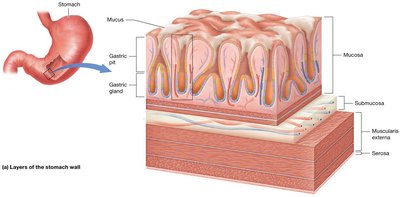
Layers: Mucosa, submucosa, muscularis externa (three layers: oblique, circular, longitudinal), serosa.
Gastric pits: Indentations in mucosa; contain columnar and goblet cells (mucus secretion).
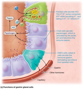
Gastric glands: At base of pits; contain mucous neck cells, parietal cells (HCl, intrinsic factor), chief cells (pepsinogen), DNES cells (hormones).
Functions of the Stomach
Secretion: HCl, pepsinogen, mucus, hormones.
Propulsion: Peristalsis and churning.
Digestion: Mechanical (churning), chemical (protein digestion by pepsin).
Acid Secretion Phases
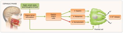
Cephalic phase: CNS stimulation (sight, smell, taste, thought of food) increases H+ secretion via vagus nerve.
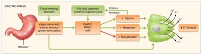
Gastric phase: Food in stomach stimulates acid secretion (distension, protein presence).
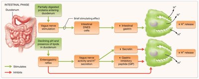
Intestinal phase: Chyme in duodenum briefly stimulates, then inhibits acid secretion (via hormones and enterogastric reflex).
Motility and Emptying
Receptive relaxation: Stomach expands to receive food.
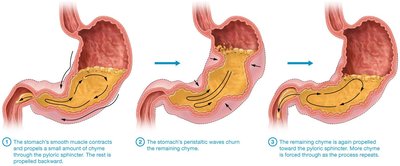
Churning: Peristaltic waves mix chyme.
Emptying: Controlled by duodenal feedback (stretch, pH, lipid content).
The Small Intestine
Divisions and Structure
Duodenum: Initial segment, receives secretions from gallbladder and pancreas.
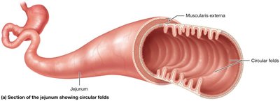
Jejunum: Middle segment, most active in digestion and absorption.
Ileum: Final segment, ends at ileocecal valve (controls flow to large intestine).
Surface Area Adaptations
Circular folds (plicae circulares): Slow transit, increase surface area.
Villi: Projections with enterocytes, capillaries, and lacteals.
Microvilli: Brush border enzymes for final digestion.
The Large Intestine
Gross Anatomy
Cecum: First portion, houses appendix.
Colon: Ascending, transverse, descending, sigmoid.
Rectum and anal canal: Terminal portions, contain internal and external sphincters.
Histology and Function
Mucosa: Lacks villi, rich in goblet cells (mucus secretion).
Muscularis externa: Longitudinal layer forms taeniae coli, creating haustra.
Serosa: Contains epiploic appendages (fat pouches).
Bacteria and Motility
Normal flora: Synthesize vitamins, metabolize undigested materials, deter pathogens, stimulate immune system.
Motility: Segmentation (churning), mass movements (peristalsis), defecation reflex.
Accessory Digestive Organs: Pancreas, Liver, Gallbladder
Pancreas
Endocrine: Insulin and glucagon.
Exocrine: Acinar cells secrete digestive enzymes; duct cells secrete bicarbonate (alkaline juice).
Pancreatic juice: Neutralizes acidic chyme, digests carbohydrates, proteins, lipids, nucleic acids.
Regulation: CCK (enzymes), secretin (bicarbonate).
Liver and Gallbladder
Liver: Four lobes, produces bile, processes nutrients, detoxifies, excretes wastes.
Liver lobule: Hexagonal unit with hepatocytes, central vein, portal triad (arteriole, venule, bile duct).
Bile: Contains bile salts (emulsify lipids), cholesterol, bilirubin, waste products.
Gallbladder: Stores and concentrates bile, releases it via cystic duct and common bile duct.
Regulation: CCK stimulates contraction and bile release; secretin stimulates bile production.
Nutrient Digestion and Absorption
General Principles
Mechanical digestion: Physical breakdown (mastication, churning, segmentation).
Chemical digestion: Enzymatic hydrolysis (water used to break bonds).
Absorption: Molecules cross enterocyte membranes; most require transport proteins.
Carbohydrate Digestion and Absorption
Salivary amylase: Begins digestion in mouth.
Pancreatic amylase: Continues in small intestine.
Brush border enzymes: Lactase, maltase, sucrase produce monosaccharides (glucose, fructose, galactose).
Protein Digestion and Absorption
Pepsin: Begins digestion in stomach.
Pancreatic enzymes: Trypsin, chymotrypsin, carboxypeptidase, etc., activated in small intestine.
Brush border enzymes: Complete digestion to amino acids.
Lipid Digestion and Absorption
Mechanical digestion: Mastication, churning, segmentation.
Bile salts: Emulsify lipids in small intestine.
Pancreatic lipase: Digests triglycerides to fatty acids and monoglycerides.
Micelles: Transport lipids to enterocytes; reassembled into triglycerides and packaged into chylomicrons for lymphatic transport.
Nucleic Acid Digestion and Absorption
Pancreatic nucleases: Break nucleic acids into nucleotides.
Brush border enzymes: Further digest to phosphate, sugar, and nitrogenous base.
Absorption: Active transport into capillaries.
Absorption of Water, Electrolytes, and Vitamins
Water: Absorbed by osmosis; most in small intestine, remainder in large intestine.
Electrolytes: Sodium, chloride, bicarbonate, iron, magnesium absorbed by various mechanisms.
Vitamins:
Water-soluble: Diffuse through membranes (except B12, which requires intrinsic factor).
Fat-soluble: Absorbed with lipids in micelles (A, D, E, K).
Summary Table: Digestive System Organs and Functions
Organ | Main Function(s) | Key Features |
|---|---|---|
Oral cavity | Ingestion, mechanical digestion | Teeth, tongue, salivary glands |
Pharynx | Propulsion | Swallowing muscles |
Esophagus | Propulsion | Sphincters, peristalsis |
Stomach | Secretion, digestion, propulsion | Gastric glands, rugae, three muscle layers |
Small intestine | Digestion, absorption, propulsion | Villi, microvilli, circular folds |
Large intestine | Absorption, propulsion, defecation | Haustra, taeniae coli, goblet cells |
Pancreas | Enzyme secretion, hormone production | Acinar cells, duct cells |
Liver | Bile production, metabolism, detoxification | Lobules, portal triad |
Gallbladder | Bile storage and concentration | Muscular sac, cystic duct |
Additional info: This guide expands on the original notes with definitions, examples, and structural context to provide a comprehensive overview suitable for exam preparation in Anatomy & Physiology.
