 Indietro
Electrical Activity and Force Generation in Cardiac Physiology
Guida di studio - Note intelligenti
 Appunti personalizzati basati sui tuoi materiali, ampliati con definizioni chiave, esempi e contesto.
Cardiac Physiology: Electrical Activity and Force Generation
Overview of Cardiac Action Potentials
The heart's rhythmic contraction is governed by specialized electrical activity in two main cell types: pacemaker cells (primarily in the SA node) and contractile cardiomyocytes. Understanding their action potentials is essential for grasping how the heart initiates and regulates its pumping function.
Pacemaker cells generate spontaneous action potentials, setting the heart rate.
Contractile cells respond to electrical signals and produce force for blood ejection.
Pacemaker Action Potentials (SA Node)
Pacemaker cells in the SA node do not maintain a stable resting membrane potential. Instead, they cycle through three main phases, enabling automatic initiation of the heartbeat.
Phase 4: Pacemaker Potential (Gradual Depolarization)
Membrane potential slowly drifts toward threshold after repolarization.
Driven by the hyperpolarization-activated "funny" current (If), reduced K+ efflux, and Ca2+ entry.
T-type Ca2+ channels open at lower thresholds, helping reach action potential threshold.
Cell is not waiting for an external signal; it approaches threshold autonomously.
Phase 0: Depolarization
Voltage-gated L-type Ca2+ channels open at threshold.
Ca2+ influx causes the rapid upstroke of the action potential.
Phase 3: Repolarization
Ca2+ channels inactivate; K+ efflux repolarizes the cell.
Membrane potential becomes more negative, restarting the pacemaker cycle.
Phase 4 Slope and Heart Rate: The steepness of phase 4 determines how quickly threshold is reached and thus the heart rate.
Sympathetic stimulation (norepinephrine, β1-adrenergic receptors): Increases cAMP, steepens phase 4, increases heart rate.
Parasympathetic stimulation (acetylcholine, M2 muscarinic receptors): Decreases cAMP, flattens phase 4, decreases heart rate.
Contractile Cell Action Potentials
Contractile cardiomyocytes exhibit a distinct action potential with a stable resting membrane potential and a prolonged plateau phase, crucial for coordinated contraction and prevention of tetanus.
Phase 4: Stable resting membrane potential.
Phase 0: Rapid Na+ influx causes depolarization.
Phase 1: Brief repolarization as Na+ channels close and L-type Ca2+ channels open.
Phase 2 (Plateau): Ca2+ influx balanced by K+ efflux.
Phase 3: K+ efflux predominates, repolarizing the cell.
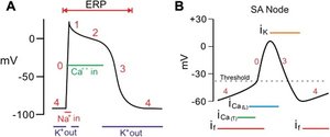
Excitation-Contraction Coupling in Cardiomyocytes
The process linking electrical activation to mechanical contraction in cardiac muscle is called excitation-contraction coupling. It involves both extracellular and intracellular calcium dynamics.
Electrical signal spreads via voltage-gated Na+, K+, and L-type Ca2+ channels.
Calcium-induced calcium release (CICR):
Action potential depolarizes the sarcolemma and T-tubules.
L-type Ca2+ channels open, allowing Ca2+ entry from extracellular space.
Incoming Ca2+ triggers ryanodine receptors in the sarcoplasmic reticulum (SR) to release more Ca2+.
Cytosolic Ca2+ rises, enabling actin-myosin cross-bridge formation and contraction.
Relaxation:
SERCA pumps return Ca2+ to the SR.
Na+/Ca2+ exchanger removes Ca2+ from the cell.
Ca2+ dissociates from troponin, tropomyosin blocks actin-myosin interaction, and force declines.
Sympathetic Regulation of Cardiac Force
Sympathetic stimulation increases cardiac contractile force (positive inotropy) by enhancing Ca2+ entry and SR Ca2+ cycling.
Norepinephrine activates β1-adrenergic receptors.
More L-type Ca2+ channels open, increasing Ca2+ influx.
SR Ca2+ cycling is enhanced, making more Ca2+ available for contraction.
Result: Greater contractile force.
Common Misconceptions in Cardiac Physiology
Misconception | Correction |
|---|---|
The action potential itself is the contraction. | Action potential triggers intracellular events leading to contraction. |
All Ca2+ needed for contraction enters from outside. | Extracellular Ca2+ triggers SR Ca2+ release. |
Ca2+ directly pulls actin and myosin together. | Ca2+ binds troponin C, allowing actin-myosin interaction. |
Relaxation happens automatically when stimulation ends. | Cytosolic Ca2+ must be removed for relaxation. |
Increased heart rate always means increased force. | Chronotropy (rate) and inotropy (force) are distinct. |
More Ca2+ always means proportionally more force. | Force depends on myofilament sensitivity and sarcomere length. |
Key Equations and Concepts
Membrane potential changes: (Nernst equation for K+ equilibrium potential)
Calcium-induced calcium release:
Practice Questions
Which event directly triggers calcium-induced calcium release in a ventricular cardiomyocyte?
A drug reduces SR Ca2+ release but does not change the ventricular action potential. What is the most likely direct effect?
Explain how sympathetic β1 stimulation increases ventricular contractile force.
Explain why Ca2+ removal is necessary for relaxation.
A patient has normal electrical activation of the ventricles, but a drug reduces Ca2+ entry through L-type channels. Explain how electrical activity can remain present while contraction becomes weaker.
Additional info: Academic context was added to clarify the phases of action potentials, excitation-contraction coupling, and the effects of autonomic regulation.
