 Indietro
Embryology and Prenatal Development: Structured Study Notes
Guida di studio - Note intelligenti
 Appunti personalizzati basati sui tuoi materiali, ampliati con definizioni chiave, esempi e contesto.
Embryology and Prenatal Development
Overview of Developmental Stages
Human development from conception to birth is divided into three main stages: pre-embryonic, embryonic, and fetal development. Each stage is characterized by distinct cellular and physiological events that lay the foundation for the formation of a viable human organism.
Pre-embryonic development: Occurs during the first two weeks after fertilization, resulting in the formation of the embryo.
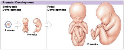
Embryonic development: Spans the third to eighth weeks, during which major organ systems begin to form.
Fetal development: Begins at the ninth week and continues until birth, focusing on growth and maturation of organ systems.
Fertilization
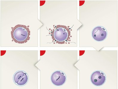
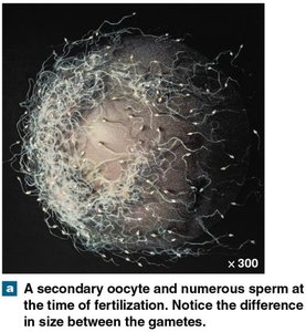
Fertilization is the process by which two haploid gametes (sperm and oocyte) fuse to form a diploid zygote. This event initiates human development and restores the chromosome number to 46.
Sperm: Delivers paternal chromosomes; highly streamlined for mobility.
Oocyte: Provides maternal chromosomes and cytoplasmic resources for early development.
Zygote: The first cell of the new organism, containing a complete set of chromosomes.
Gestation and Trimesters
Gestation is the period of prenatal development, divided into three trimesters, each lasting approximately three months. Each trimester is marked by specific developmental milestones.
First trimester: Includes cleavage, implantation, placentation, and embryogenesis. Rudiments of all major organ systems appear.
Second trimester: Organ systems develop further; body shape and proportions change.
Third trimester: Rapid fetal growth, adipose tissue deposition, and functional maturation of organ systems.
First Trimester Processes
Cleavage
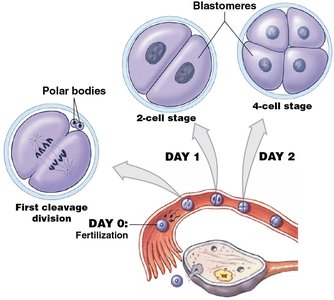
Cleavage is a series of rapid mitotic divisions that transform the zygote into a multicellular blastocyst. This process begins immediately after fertilization and ends when the blastocyst contacts the uterine wall.
Blastomeres: The cells produced by cleavage divisions.
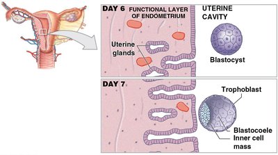
Blastocyst: A hollow ball of cells that will implant in the uterine wall.
Implantation
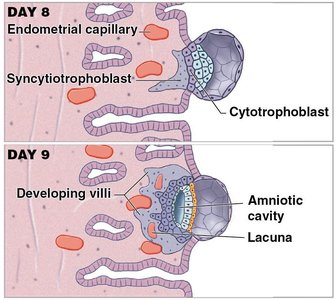
Implantation is the attachment of the blastocyst to the endometrium of the uterus, occurring 7–10 days after fertilization. Trophoblast cells facilitate implantation by secreting enzymes that erode the uterine epithelium.
Cytotrophoblast: Inner layer of trophoblast cells.
Syncytiotrophoblast: Outer layer that invades the endometrium.
Placentation
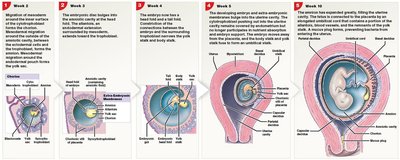
Placentation involves the formation of the placenta, which permits exchange of nutrients, gases, and wastes between maternal and embryonic blood. Blood vessels form around the blastocyst, and the umbilical cord connects the fetus to the placenta.
Body stalk: Connects embryo to chorion; contains allantois and blood vessels.
Yolk stalk: Connects endoderm of embryo to yolk sac.
Umbilical arteries and vein: Facilitate placental circulation.
Embryogenesis
Embryogenesis is the formation of a viable embryo and the establishment of the foundations for all major organ systems. Gastrulation forms three primary germ layers: ectoderm, mesoderm, and endoderm.
Ectoderm: Forms skin and nervous system.
Mesoderm: Forms muscles, bones, and circulatory system.
Endoderm: Forms digestive and respiratory systems.
Second and Third Trimesters
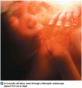
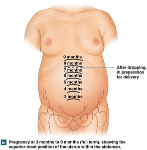
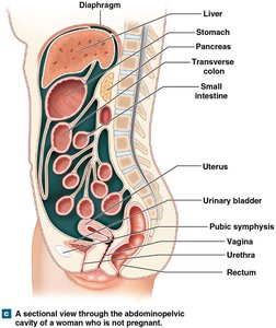
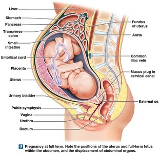
During the second trimester, the fetus grows rapidly and the amnion and chorion fuse to form the amniochorionic membrane. In the third trimester, organ systems mature and the fetus gains significant weight, displacing maternal abdominal organs.
Maternal Adaptations During Pregnancy
The developing fetus relies entirely on maternal organ systems for nourishment, respiration, and waste removal. Pregnancy induces several physiological adaptations in the mother.
Respiratory rate and tidal volume: Increase to meet fetal oxygen demands.
Blood volume: Increases by almost 50%.
Nutrient intake: Increases by 10–30%.
Glomerular filtration rate: Increases by 50% for waste removal.
Uterus and mammary glands: Increase in size.
Growth of Uterus and Fetus During Gestation
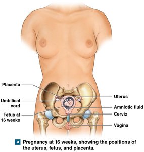
As gestation progresses, the uterus expands to accommodate the growing fetus, and the positions of maternal organs shift accordingly. The fetus is surrounded by amniotic fluid and connected to the placenta via the umbilical cord.
Summary Table: Key Events in Early Development
Day/Week | Event | Description |
|---|---|---|
Day 0 | Fertilization | Fusion of sperm and oocyte; formation of zygote |
Day 1-2 | Cleavage | Mitotic divisions; formation of blastomeres |
Day 3-4 | Morula | Solid ball of cells; zona pellucida present |
Day 6-10 | Blastocyst & Implantation | Blastocyst contacts and adheres to uterine wall |
Week 3-8 | Embryonic development | Formation of organ systems; gastrulation |
Week 9-birth | Fetal development | Growth and maturation of organ systems |
Key Terms and Definitions
Gestation: The period of prenatal development from conception to birth.
Blastocyst: A hollow ball of cells formed during early embryonic development.
Placenta: Organ that facilitates exchange between maternal and fetal blood.
Umbilical cord: Structure connecting fetus to placenta, containing blood vessels.
Germ layers: Primary layers (ectoderm, mesoderm, endoderm) formed during gastrulation.
Relevant Equations
While embryology is primarily descriptive, the following formula is relevant for chromosome number:
Chromosome number after fertilization:
Additional info: Academic context was added to clarify the stages of development, the role of maternal adaptations, and the significance of key terms for exam preparation.
