 Indietro
Endocrine and Cardiovascular Systems: Study Guide (Chapters 16-18)
Guida di studio - Note intelligenti
 Appunti personalizzati basati sui tuoi materiali, ampliati con definizioni chiave, esempi e contesto.
Endocrine System
Hormones of the Adrenal Gland and Their Functions
The adrenal glands are located above the kidneys and secrete several important hormones that regulate metabolism, stress response, and electrolyte balance.
Cortex Hormones:
Mineralocorticoids (e.g., aldosterone): Regulate sodium and potassium balance, affecting blood pressure.
Glucocorticoids (e.g., cortisol): Influence metabolism, immune response, and stress adaptation.
Androgens: Contribute to secondary sex characteristics.
Medulla Hormones:
Epinephrine (adrenaline) and norepinephrine: Mediate the fight-or-flight response, increasing heart rate and blood glucose.
Anatomical and Physiological Connections of the Hypothalamus and Pituitary Glands
The hypothalamus and pituitary gland form a major regulatory axis in the endocrine system.
Anatomical Connection: The pituitary gland is attached to the hypothalamus by the infundibulum.
Physiological Connection: The hypothalamus produces releasing and inhibiting hormones that control the anterior pituitary. The posterior pituitary stores and releases hormones produced by the hypothalamus (e.g., ADH, oxytocin).
Functional Classifications of Hormones
Hormones can be classified based on their chemical structure and mechanism of action.
Amino acid-based hormones: Includes peptides, proteins, and amines (e.g., insulin, epinephrine).
Steroid hormones: Derived from cholesterol (e.g., cortisol, aldosterone, sex hormones).
Mechanism of Action: Water-soluble hormones act via membrane receptors; lipid-soluble hormones act via intracellular receptors.
Hormones of Calcium Homeostasis
Calcium homeostasis is regulated by three main hormones:
Parathyroid hormone (PTH): Increases blood calcium by stimulating bone resorption, increasing intestinal absorption, and reducing renal excretion.
Calcitonin: Lowers blood calcium by inhibiting bone resorption.
Calcitriol (active vitamin D): Increases intestinal absorption of calcium.
Thymus Gland
The thymus is located in the mediastinum and is most active during childhood. It produces thymosins, which are essential for T-lymphocyte maturation and immune function.
Homeostasis of Blood Sugar
Blood glucose is regulated primarily by the pancreas:
Insulin: Lowers blood glucose by promoting cellular uptake and storage as glycogen.
Glucagon: Raises blood glucose by stimulating glycogen breakdown and gluconeogenesis.
Cardiovascular System
The Heart Anatomy, Including Conduction System
The heart is a muscular organ with four chambers (two atria, two ventricles) and a specialized conduction system that coordinates contraction.
Conduction System: Includes the sinoatrial (SA) node, atrioventricular (AV) node, bundle of His, bundle branches, and Purkinje fibers.
Function: Ensures rhythmic and coordinated contraction of the heart chambers.
Blood Flow in the Chambers of the Heart
Blood flows through the heart in a specific sequence:
Right atrium → right ventricle → pulmonary artery → lungs
Lungs → pulmonary veins → left atrium → left ventricle → aorta → body
Anatomy of the Walls of the Heart
The heart wall consists of three layers:
Epicardium: Outer layer (visceral pericardium)
Myocardium: Middle, muscular layer responsible for contraction
Endocardium: Inner endothelial lining
Coronary Cardiac Circuit Anatomy
The coronary circulation supplies blood to the heart muscle itself via the right and left coronary arteries, cardiac veins, and the coronary sinus.
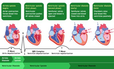
Cardiac Cycle Events – 4 Phases
The cardiac cycle consists of a sequence of events that occur during one heartbeat:
Atrial systole: Atria contract, pushing blood into ventricles.
Ventricular systole (first phase): Ventricles contract, AV valves close.
Ventricular systole (second phase): Semilunar valves open, blood is ejected.
Ventricular diastole: Ventricles relax, semilunar valves close, chambers fill passively.
Pulmonary and Systemic Blood Circulation
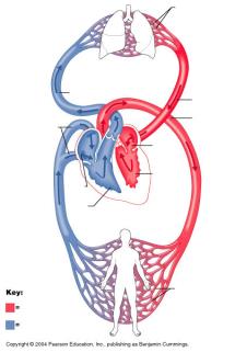
The heart pumps blood through two main circuits:
Pulmonary circuit: Right side of the heart pumps deoxygenated blood to the lungs for oxygenation.
Systemic circuit: Left side of the heart pumps oxygenated blood to the rest of the body.
Cardiac Output Law
Cardiac output (CO) is the volume of blood pumped by each ventricle per minute.
Formula:
Where HR is heart rate and SV is stroke volume.
Stroke Volume Laws
Stroke volume (SV) is influenced by preload, afterload, and contractility.
Preload: Degree of stretch of cardiac muscle fibers at the end of diastole.
Afterload: Resistance the ventricles must overcome to eject blood.
Contractility: Strength of ventricular contraction.
ECG: All Peaks, Segments, and Intervals
An electrocardiogram (ECG) records the electrical activity of the heart:
P wave: Atrial depolarization
QRS complex: Ventricular depolarization
T wave: Ventricular repolarization
PR interval: Time from atrial to ventricular depolarization
ST segment: Time between ventricular depolarization and repolarization
Sinus Rhythm
Sinus rhythm is the normal, regular rhythm of the heart initiated by the SA node, typically 60-100 beats per minute in adults.
Sounds of the Heart
The heart produces two main sounds:
"Lub" (S1): Closure of AV valves at the beginning of ventricular systole.
"Dub" (S2): Closure of semilunar valves at the beginning of ventricular diastole.
Athlete ESV, EDV
End-systolic volume (ESV) and end-diastolic volume (EDV) are typically lower and higher, respectively, in athletes due to increased cardiac efficiency and chamber size.
EDV: Volume of blood in ventricle at end of filling (diastole).
ESV: Volume of blood remaining after contraction (systole).
Anatomy of the Blood Vessels
Blood vessels include arteries, veins, and capillaries, each with distinct structural features:
Arteries: Thick, muscular walls to withstand high pressure.
Veins: Thinner walls, valves to prevent backflow.
Capillaries: Thin walls for exchange of gases, nutrients, and wastes.
Blood Pressure Forces Components
Blood pressure is determined by cardiac output, blood volume, and resistance of blood vessels.
Systolic pressure: Pressure during ventricular contraction.
Diastolic pressure: Pressure during ventricular relaxation.
Blood Pressure Modifying Factors
Cardiac output
Blood volume
Peripheral resistance
Elasticity of vessel walls
Blood Pressure Variation Throughout the Body
Blood pressure is highest in the arteries and decreases through arterioles, capillaries, venules, and veins.
Role of SNS and PSNS in BP Regulation
The sympathetic nervous system (SNS) increases heart rate and vasoconstriction, raising blood pressure. The parasympathetic nervous system (PSNS) decreases heart rate, lowering blood pressure.
Mean Arterial Pressure
Mean arterial pressure (MAP) is the average pressure in the arteries during one cardiac cycle.
Formula:
Types of Capillaries
Continuous: Most common, uninterrupted lining.
Fenestrated: Pores for increased permeability (e.g., kidneys, intestines).
Sinusoidal: Large gaps for passage of large molecules (e.g., liver, spleen).
Mechanisms of Blood Pressure Regulation
Short-term: Neural (baroreceptors), hormonal (epinephrine, ADH), and local mechanisms.
Long-term: Renal regulation of blood volume.
Capillaries Net Filtration Factors
Net filtration at capillaries is determined by hydrostatic and osmotic pressures:
Net Filtration Pressure (NFP):
Where HP = hydrostatic pressure, OP = osmotic pressure, c = capillary, i = interstitial fluid.
Upper Limb Arteries
Major arteries of the upper limb include the subclavian, axillary, brachial, radial, and ulnar arteries.
Major Veins
Major veins include the superior and inferior vena cava, jugular veins, subclavian veins, and femoral veins.
Case Study 1: Thyroid Gland Disorders
Thyroid disorders affect hormone levels and feedback mechanisms:
Hypothyroidism: Low T3/T4, high TSH (if primary), due to lack of negative feedback.
Hyperthyroidism: High T3/T4, low TSH (if primary), due to excessive negative feedback.
Feedback System: Hypothalamus releases TRH → pituitary releases TSH → thyroid releases T3/T4; negative feedback regulates TRH and TSH.
Case Study 2: Heart Interventricular Defected Wall
An interventricular septal defect allows blood to flow between the ventricles, affecting stroke volume (SV) and EKG:
Stroke Volume: May decrease due to inefficient pumping and mixing of oxygenated/deoxygenated blood.
EKG: May show abnormal QRS complexes or other conduction disturbances.
