 Indietro
Fundamentals of the Nervous System and Nervous Tissue
Guida di studio - Note intelligenti
 Appunti personalizzati basati sui tuoi materiali, ampliati con definizioni chiave, esempi e contesto.
Fundamentals of the Nervous System and Nervous Tissue
Overview of the Nervous System
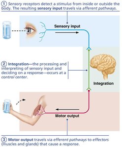
The nervous system is the master controlling and communicating system of the body. It utilizes both electrical and chemical signals to coordinate rapid and specific responses, often resulting in immediate effects. The nervous system is responsible for sensory input, integration, and motor output.
Sensory Input: Information gathered by sensory receptors about internal and external changes.
Integration: Processing and interpretation of sensory input to determine an appropriate response.
Motor Output: Activation of effectors (muscles and glands) to produce a response.
Subdivisions of the Nervous System
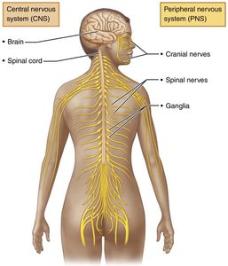
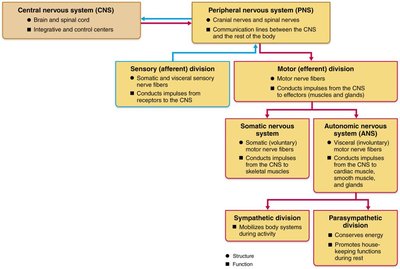
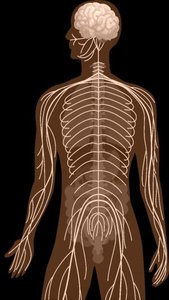
The nervous system is divided into two principal parts: the Central Nervous System (CNS) and the Peripheral Nervous System (PNS).
Central Nervous System (CNS): Consists of the brain and spinal cord, serving as the integration and control center. It interprets sensory input and dictates motor output.
Peripheral Nervous System (PNS): Composed mainly of nerves that extend from the brain and spinal cord (cranial and spinal nerves). It connects the CNS to limbs and organs.
Functional Divisions of the PNS
Sensory (Afferent) Division: Transmits impulses from sensory receptors to the CNS.
Somatic sensory fibers: Convey impulses from skin, skeletal muscles, and joints.
Visceral sensory fibers: Convey impulses from visceral organs.
Motor (Efferent) Division: Transmits impulses from the CNS to effector organs (muscles and glands).
Somatic Nervous System: Voluntary control of skeletal muscles.
Autonomic Nervous System (ANS): Involuntary control of smooth muscle, cardiac muscle, and glands.
Sympathetic Division: Mobilizes body systems during activity.
Parasympathetic Division: Conserves energy and promotes housekeeping functions during rest.
Cellular Components of Nervous Tissue
Main Types of Cells
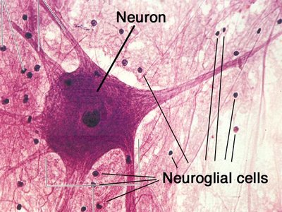
Nervous tissue consists of two principal cell types:
Neuroglia (Glial Cells): Small cells that support, protect, and insulate neurons.
Neurons (Nerve Cells): Excitable cells that transmit electrical signals.
Neuroglia of the CNS
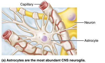
Astrocytes: Most abundant glial cells; support neurons, regulate the chemical environment, and facilitate nutrient exchange between capillaries and neurons.
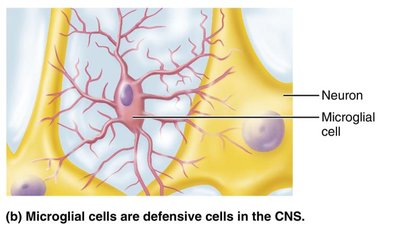
Microglial Cells: Small, defensive cells that monitor neuron health and can transform into phagocytes to remove debris and pathogens.
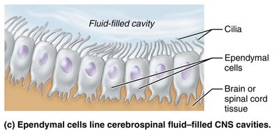
Ependymal Cells: Line the central cavities of the brain and spinal cord; may be ciliated to help circulate cerebrospinal fluid (CSF).
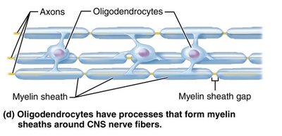
Oligodendrocytes: Form myelin sheaths around CNS nerve fibers, insulating them and increasing the speed of impulse transmission.
Neuroglia of the PNS
Satellite Cells: Surround neuron cell bodies in the PNS, providing support and regulating the environment.
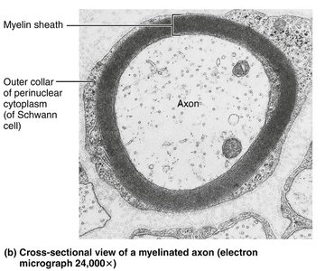
Schwann Cells (Neurolemmocytes): Surround all peripheral nerve fibers and form myelin sheaths around thicker fibers; vital for regeneration of damaged peripheral nerve fibers.
Neurons: Structure and Function
Main Parts of a Neuron
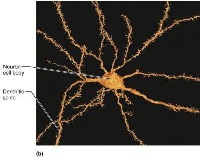
Cell Body (Soma): Contains the nucleus and cytoplasm; receptive region for incoming signals. Most neuron cell bodies are located in the CNS (called nuclei); in the PNS, they are found in ganglia.
Processes: Extensions from the cell body, including dendrites and axons.
Dendrites: Short, branched processes that receive input and convey it toward the cell body as graded potentials.
Axon: Each neuron has a single axon that starts at the axon hillock and transmits impulses away from the cell body. Axons may be long (nerve fibers) and branch at their ends (axon terminals), which release neurotransmitters to communicate with other cells.
Myelin Sheath
The myelin sheath is a white, fatty covering that insulates axons, increasing the speed of nerve impulse transmission. Myelinated axons conduct impulses faster than nonmyelinated axons. Myelin is produced by Schwann cells in the PNS and oligodendrocytes in the CNS.
Nodes of Ranvier: Gaps between adjacent Schwann cells (PNS) or oligodendrocyte processes (CNS) where axon membrane is exposed; important for rapid signal conduction.
Classification of Neurons
Structural Classification
Multipolar Neurons: Three or more processes (one axon, multiple dendrites); most common in the CNS.
Bipolar Neurons: Two processes (one axon, one dendrite); rare, found in special sense organs (e.g., retina, olfactory mucosa).
Unipolar (Pseudounipolar) Neurons: Single short process that splits into two branches; primarily sensory neurons in the PNS.
Functional Classification
Sensory (Afferent) Neurons: Transmit impulses from sensory receptors toward the CNS; mostly unipolar, with cell bodies in PNS ganglia.
Motor (Efferent) Neurons: Carry impulses from the CNS to effectors (muscles/glands); mostly multipolar, with cell bodies in the CNS.
Interneurons (Association Neurons): Lie between sensory and motor neurons within the CNS; responsible for integration and signal processing; make up 99% of all neurons.
Summary Table: Structural and Functional Classification of Neurons
Structural Type | Main Location | Function |
|---|---|---|
Multipolar | CNS | Motor neurons, interneurons |
Bipolar | Special sense organs (retina, olfactory mucosa) | Sensory neurons for special senses |
Unipolar | PNS (dorsal root ganglia) | Primary sensory neurons |
Example: A multipolar motor neuron conducts impulses from the spinal cord to skeletal muscle, while a unipolar sensory neuron transmits impulses from the skin to the spinal cord.
Additional info: The enteric nervous system, found in the walls of the gastrointestinal tract, is sometimes considered a third division of the nervous system due to its autonomy and complexity.
