 Indietro
Fundamentals of the Nervous System and Nervous Tissue: Study Notes
Guida di studio - Note intelligenti
 Appunti personalizzati basati sui tuoi materiali, ampliati con definizioni chiave, esempi e contesto.
Fundamentals of the Nervous System and Nervous Tissue
Overview of the Nervous System
The nervous system is the master controlling and communicating system of the body. It is designed to receive input from both the external and internal environments, integrate this information, and coordinate appropriate responses. The nervous system maintains homeostasis by detecting changes, making decisions, and stimulating muscles and glands to respond. Communication within the nervous system occurs via rapid and specific electrical and chemical signals.
Sensory Input: Information gathered by sensory receptors about internal and external changes, transmitted to the CNS via afferent (sensory) neurons.
Integration: Processing and interpretation of sensory input by interneurons within the CNS.
Motor Output: Activation of effector organs (muscles and glands) to produce a response via motor (efferent) neurons.
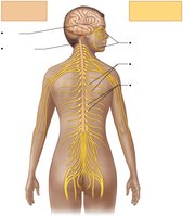
Divisions of the Nervous System
Central Nervous System (CNS): Consists of the brain and spinal cord. It is the integration and control center, interpreting sensory input and dictating motor output.
Peripheral Nervous System (PNS): Consists mainly of nerves that extend from the brain and spinal cord (cranial and spinal nerves). It serves as communication lines between the CNS and the rest of the body.
Functional Divisions of the PNS
Sensory (Afferent) Division: Transmits impulses from sensory receptors to the CNS. Includes somatic sensory fibers (from skin, skeletal muscles, joints) and visceral sensory fibers (from visceral organs).
Motor (Efferent) Division: Transmits impulses from the CNS to effector organs. Subdivided into:
Somatic Nervous System: Voluntary control of skeletal muscles.
Autonomic Nervous System (ANS): Involuntary control of smooth muscle, cardiac muscle, and glands. Further divided into sympathetic (mobilizes body systems during activity) and parasympathetic (promotes housekeeping functions during rest) divisions.
Histology of Nervous Tissue
Cell Types in Nervous Tissue
Nervous tissue is highly cellular, with little extracellular space. It consists of two principal cell types:
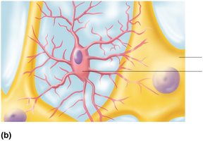
Neurons (Nerve Cells): Excitable cells that transmit electrical signals. They are the structural and functional units of the nervous system.
Neuroglia (Glial Cells): Supporting cells that surround and wrap neurons. They outnumber neurons by about 10:1 and perform various supportive functions.
Neuroglia of the CNS
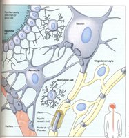
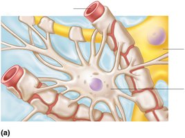
Astrocytes: Most abundant, versatile, and highly branched. They support and brace neurons, help maintain the blood-brain barrier, and participate in information processing.
Microglial Cells: Small, ovoid cells that act as the CNS immune defense by phagocytizing microorganisms and debris.
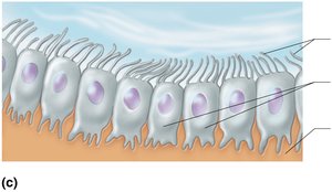
Ependymal Cells: Line the central cavities of the brain and spinal cord; may be ciliated to help circulate cerebrospinal fluid (CSF).
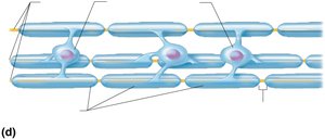
Oligodendrocytes: Form myelin sheaths around CNS nerve fibers, insulating them and increasing conduction speed.
Neuroglia of the PNS
Satellite Cells: Surround neuron cell bodies in the PNS, providing support and nourishment.
Schwann Cells (Neurolemmocytes): Surround all peripheral nerve fibers and form myelin sheaths in the PNS. Vital for regeneration of damaged peripheral nerve fibers.
Neurons: Structure and Function
General Structure of Neurons
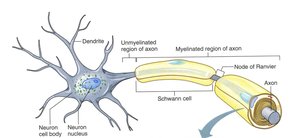
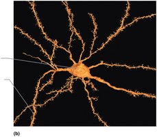
Neurons are large, highly specialized cells that conduct impulses. They have extreme longevity, are mostly amitotic, and have a high metabolic rate. All neurons have a cell body (soma) and one or more processes (dendrites and axons).
Cell Body (Soma): Contains the nucleus and organelles; biosynthetic center of the neuron.
Dendrites: Short, branched extensions that receive input and convey it toward the cell body.
Axon: Long process that transmits impulses away from the cell body to other neurons or effectors. Axons may be myelinated or unmyelinated.
Myelin Sheath
The myelin sheath is a segmented, lipid-rich covering around most long or large-diameter axons. It protects and electrically insulates axons, increasing the speed of nerve impulse transmission.
In the PNS: Formed by Schwann cells wrapping around the axon in a jelly roll fashion.
In the CNS: Formed by oligodendrocytes, each of which can myelinate multiple axons.
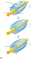
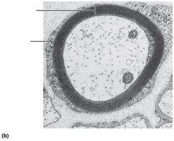
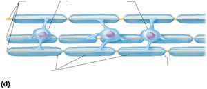
White Matter vs. Gray Matter
White Matter: Regions with dense collections of myelinated fibers (mainly fiber tracts).
Gray Matter: Mostly neuron cell bodies and nonmyelinated fibers (cortex and nuclei of brain, horns of spinal cord).
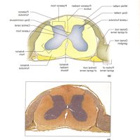
Classification of Neurons
Structural Classification
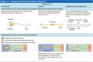
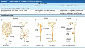
Multipolar: Three or more processes (one axon, multiple dendrites); most common in CNS.
Bipolar: Two processes (one axon, one dendrite); rare, found in retina and olfactory mucosa.
Unipolar (Pseudounipolar): One T-like process; found mainly in PNS sensory neurons.
Functional Classification
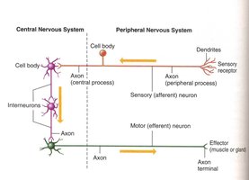
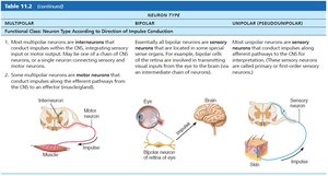
Sensory (Afferent) Neurons: Transmit impulses from sensory receptors toward the CNS; mostly unipolar.
Motor (Efferent) Neurons: Carry impulses from the CNS to effectors; multipolar.
Interneurons (Association Neurons): Shuttle signals within the CNS; most are multipolar and make up 99% of neurons.
Membrane Potentials and Electrical Signals
Basic Principles of Electricity
Voltage (V): Measure of potential energy generated by separated charge; measured in volts (V) or millivolts (mV).
Current (I): Flow of electrical charge (ions) between two points.
Resistance (R): Hindrance to charge flow; insulators have high resistance, conductors have low resistance.
Ohm’s Law:
Ion Channels
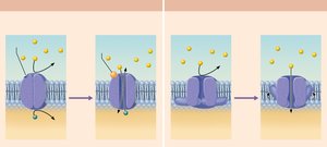
Chemically Gated (Ligand-Gated) Channels: Open with binding of a specific chemical (e.g., neurotransmitter).
Voltage-Gated Channels: Open and close in response to changes in membrane potential.
Mechanically Gated Channels: Open in response to physical deformation of receptors.
Resting Membrane Potential
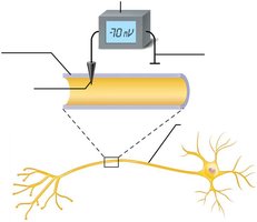
Neurons have a resting membrane potential (RMP), typically around -70 mV. This is maintained by differences in ion concentrations across the membrane and selective permeability of the plasma membrane to these ions. The sodium-potassium pump (Na+/K+ ATPase) helps maintain these gradients by pumping 3 Na+ out and 2 K+ in.
Changes in Membrane Potential
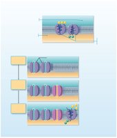
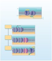
Depolarization: Decrease in membrane potential (inside becomes less negative); increases probability of producing an impulse.
Hyperpolarization: Increase in membrane potential (inside becomes more negative); decreases probability of producing an impulse.
Graded Potentials and Action Potentials
Graded Potentials
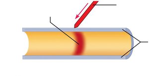
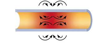
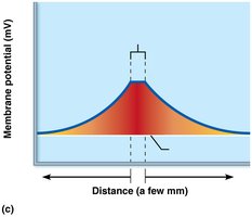
Graded potentials are short-lived, localized changes in membrane potential. They occur in dendrites and cell bodies, and their magnitude varies with stimulus strength. Graded potentials decay with distance due to leaky plasma membranes.
Action Potentials
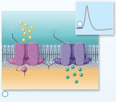
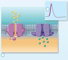
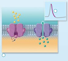
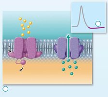
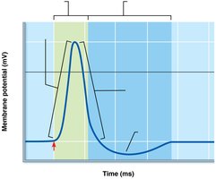
Action potentials (APs) are the principal means of long-distance neural communication. They are brief reversals of membrane potential (~100 mV) and do not decay over distance. APs are generated by the opening of voltage-gated Na+ and K+ channels.
Resting State: All gated Na+ and K+ channels are closed.
Depolarization: Na+ channels open, Na+ enters the cell.
Repolarization: Na+ channels inactivate, K+ channels open, K+ exits the cell.
Hyperpolarization: Some K+ channels remain open, Na+ channels reset.
Threshold and All-or-None Principle
For an AP to occur, depolarization must reach a threshold voltage. If threshold is reached, the AP is generated completely; if not, no AP occurs. This is known as the all-or-none phenomenon.
Propagation of Action Potentials
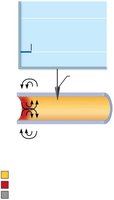
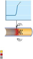
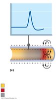
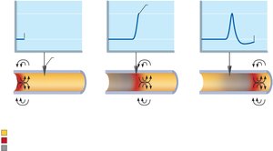
APs propagate along the axon by opening voltage-gated Na+ channels in adjacent membrane areas. In myelinated axons, APs jump from node to node (saltatory conduction), which is much faster than continuous conduction in unmyelinated axons.
Coding for Stimulus Intensity
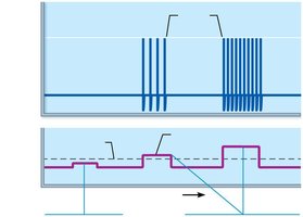
All APs are alike; the CNS distinguishes stimulus intensity by the frequency of APs. Stronger stimuli generate APs more frequently.
Refractory Periods
Absolute Refractory Period: Time during which another AP cannot be generated, regardless of stimulus strength.
Relative Refractory Period: Time during which a stronger-than-usual stimulus is required to generate another AP.
Conduction Velocity
Axon Diameter: Larger diameter fibers conduct impulses faster.
Degree of Myelination: Myelinated fibers conduct impulses much faster (saltatory conduction) than unmyelinated fibers (continuous conduction).
Types of Nerve Fibers
Group | Diameter | Myelination | Speed | Function |
|---|---|---|---|---|
A | Largest | Myelinated | 150 m/s | Somatic sensory & motor fibers |
B | Intermediate | Lightly myelinated | 15 m/s | ANS visceral motor & sensory fibers |
C | Smallest | Unmyelinated | 1 m/s | ANS visceral motor & sensory fibers |
Synapses and Neurotransmission
The Synapse
Synapses are junctions that mediate information transfer from one neuron to another or to an effector cell. The presynaptic neuron sends the signal, and the postsynaptic neuron receives it. Synapses can be axodendritic, axosomatic, axoaxonal, dendrodendritic, or somatodendritic.
Chemical Synapses
Most common type; involve the release of neurotransmitters from the presynaptic neuron, which diffuse across the synaptic cleft and bind to receptors on the postsynaptic neuron, generating graded potentials.
AP arrives at axon terminal.
Voltage-gated Ca2+ channels open; Ca2+ enters axon terminal.
Ca2+ entry causes synaptic vesicles to release neurotransmitter by exocytosis.
Neurotransmitter diffuses across synaptic cleft and binds to receptors on postsynaptic membrane.
Binding opens ion channels, resulting in graded potentials.
Neurotransmitter effects are terminated by reuptake, enzymatic degradation, or diffusion away.
Electrical Synapses
Less common; neurons are electrically coupled via gap junctions, allowing rapid, bidirectional communication. Found in some brain regions and embryonic nervous tissue.
Postsynaptic Potentials
Excitatory Postsynaptic Potentials (EPSPs): Local depolarizations that bring the neuron closer to AP threshold.
Inhibitory Postsynaptic Potentials (IPSPs): Local hyperpolarizations that drive the neuron away from AP threshold.
Summation
Temporal Summation: Multiple EPSPs from one presynaptic neuron in rapid succession add together.
Spatial Summation: EPSPs from multiple presynaptic neurons occur simultaneously and add together.
IPSPs can also summate and can counteract EPSPs.
Neurotransmitters
Classification by Chemical Structure
Acetylcholine (ACh): First identified; released at neuromuscular junctions and by many ANS neurons.
Biogenic Amines: Catecholamines (dopamine, norepinephrine, epinephrine) and indolamines (serotonin, histamine).
Amino Acids: Glutamate, aspartate, glycine, GABA.
Peptides (Neuropeptides): Substance P, endorphins, enkephalins, somatostatin, cholecystokinin.
Purines: ATP, adenosine.
Gases and Lipids: Nitric oxide (NO), carbon monoxide (CO), hydrogen sulfide (H2S), endocannabinoids.
Classification by Function
Effects: Excitatory (depolarizing) or inhibitory (hyperpolarizing), depending on receptor type.
Actions: Direct (binds and opens ion channels) or indirect (acts through second messengers, e.g., G protein pathways).
Neurotransmitter Receptors
Channel-Linked Receptors: Ligand-gated ion channels; mediate fast synaptic transmission.
G Protein–Linked Receptors: Mediate slow, prolonged responses via second messengers (e.g., cAMP).
Neural Integration and Circuits
Neuronal Pools
Neuronal pools are functional groups of neurons that integrate incoming information and forward processed information to other destinations. They have a discharge zone (neurons most likely to fire) and a facilitated zone (neurons less likely to fire).
Patterns of Neural Processing
Serial Processing: Input travels along one pathway to a specific destination (e.g., reflex arcs).
Parallel Processing: Input travels along several pathways, allowing multiple responses (important for higher-level mental functioning).
Types of Circuits
Diverging Circuit: One input, many outputs (amplifying circuit).
Converging Circuit: Many inputs, one output (concentrating circuit).
Reverberating Circuit: Signal travels through a chain of neurons, each feeding back to previous neurons (oscillating circuit).
Parallel After-Discharge Circuit: Signal stimulates neurons arranged in parallel arrays that converge on a single output cell.
Developmental Aspects of Neurons
Nervous system originates from the neural tube and neural crest (ectoderm).
Neuroblasts become amitotic and migrate, sprouting axons to connect with targets.
Growth cones guide axons to targets using adhesion proteins and neurotropins.
Astrocytes provide support and cholesterol for synapse construction.
Many neurons die before birth if they do not form synapses; apoptosis is common.
Learning during childhood and adolescence reinforces certain synapses and prunes others.
Additional info: This guide covers the essential concepts of nervous system structure, function, and cellular mechanisms, providing a foundation for further study in neurophysiology and related clinical applications.
