 Indietro
Fundamentals of the Nervous System and Nervous Tissue: Structured Study Notes
Guida di studio - Note intelligenti
 Appunti personalizzati basati sui tuoi materiali, ampliati con definizioni chiave, esempi e contesto.
Fundamentals of the Nervous System and Nervous Tissue
Overview of the Nervous System
The nervous system is the master controlling and communicating system of the body. It utilizes electrical and chemical signals to coordinate rapid, specific responses. The nervous system is divided into two principal parts: the Central Nervous System (CNS) and the Peripheral Nervous System (PNS).
Central Nervous System (CNS): Consists of the brain and spinal cord. It serves as the integration and control center, interpreting sensory input and dictating motor output.
Peripheral Nervous System (PNS): Composed of nerves extending from the brain and spinal cord, including cranial and spinal nerves. It connects the CNS to the rest of the body.
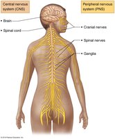
Functions of the Nervous System
The nervous system performs three overlapping functions:
Sensory Input: Gathering information from sensory receptors about internal and external changes.
Integration: Processing and interpretation of sensory input.
Motor Output: Activation of effector organs (muscles and glands) to produce a response.
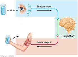
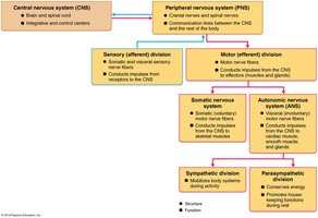
Organization of the Nervous System
The PNS is further divided into functional divisions:
Sensory (Afferent) Division: Transmits sensory information to the CNS.
Motor (Efferent) Division: Transmits impulses from the CNS to effector organs.
The motor division is subdivided into:
Somatic Nervous System: Controls voluntary movements of skeletal muscles.
Autonomic Nervous System (ANS): Regulates involuntary functions (smooth muscle, cardiac muscle, glands) and is further divided into:
Sympathetic Division: Mobilizes body systems during activity (fight or flight).
Parasympathetic Division: Conserves energy and promotes housekeeping functions (rest and digest).
Neuroglia and Neurons
Neuroglia (Glial Cells)
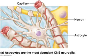
Nervous tissue consists of two principal cell types: neuroglia and neurons. Neuroglia are supporting cells that surround and wrap neurons. Four main types support CNS neurons:
Astrocytes: Most abundant, versatile, and highly branched. They support and brace neurons, facilitate exchanges between capillaries and neurons, and guide migration of young neurons.
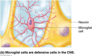
Microglial Cells: Small, defensive cells that monitor neurons and can transform to phagocytize microorganisms and debris.
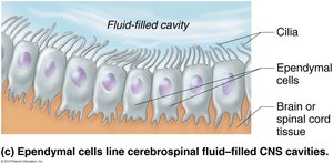
Ependymal Cells: Line cerebrospinal fluid-filled CNS cavities; their cilia help circulate CSF.
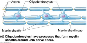
Oligodendrocytes: Branched cells that form myelin sheaths around CNS nerve fibers.
Neuroglia of the PNS
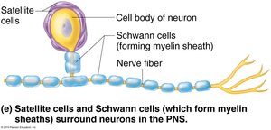
Satellite Cells: Surround neuron cell bodies in the PNS, similar in function to astrocytes.
Schwann Cells: Form myelin sheaths around peripheral nerve fibers and are vital for regeneration of damaged PNS fibers.
Neurons (Nerve Cells)
Neurons are the structural units of the nervous system, specialized for conducting impulses. They exhibit extreme longevity, are mostly amitotic, and have a high metabolic rate. Each neuron consists of a cell body and one or more processes.
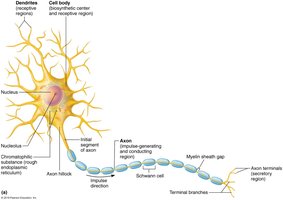
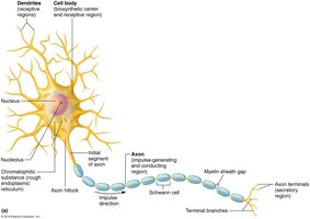
Cell Body (Soma/Perikaryon): Biosynthetic center, contains nucleus, rough ER (Nissl bodies), and is the receptive region for input.
Processes: Dendrites (receptive/input region) and axons (impulse-generating and conducting region).
Dendrites
Short, highly branched processes that receive signals and convey them toward the cell body as graded potentials.
Contain dendritic spines for specialized information collection.
Axons
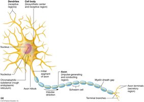
Each neuron has one axon, which may be very long (nerve fiber).
Axons branch at their ends (axon terminals) and are the site of neurotransmitter release.
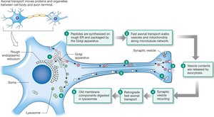
Axons transport molecules and organelles via anterograde (away from cell body) and retrograde (toward cell body) transport.
Myelination and Neuron Classification
Myelin Sheath
The myelin sheath is a protein-lipid layer that insulates axons and increases the speed of nerve impulse transmission. Myelinated fibers conduct impulses faster than nonmyelinated fibers.
CNS: Myelin formed by oligodendrocytes; white matter consists of myelinated fibers, gray matter of cell bodies and nonmyelinated fibers.
PNS: Myelin formed by Schwann cells; myelin sheath gaps (nodes of Ranvier) are sites for impulse propagation.
Classification of Neurons
Structural Classification
Multipolar: Three or more processes (one axon, others dendrites); most common in CNS.
Bipolar: Two processes (one axon, one dendrite); rare, found in retina and olfactory mucosa.
Unipolar: One T-like process (two axons); mostly sensory neurons.
Functional Classification
Sensory (Afferent): Transmit impulses toward CNS; mostly unipolar.
Motor (Efferent): Carry impulses from CNS to effectors; multipolar.
Interneurons: Shuttle signals within CNS; most abundant.
Membrane Potentials and Electrical Signaling
Basic Principles of Electricity
Neurons have a resting membrane potential and can rapidly change it. Key terms:
Voltage: Potential energy from separated charges, measured in millivolts (mV).
Current: Flow of electrical charge (ions) between two points.
Role of Membrane Ion Channels
Leakage Channels: Always open.
Gated Channels: Open/close in response to stimuli (chemical, voltage, mechanical).
Resting Membrane Potential
The resting membrane potential is maintained by ion gradients and selective permeability. Typical value is around -70 mV.
Changes in Membrane Potential
Depolarization: Membrane potential becomes less negative; increases likelihood of action potential.
Hyperpolarization: Membrane potential becomes more negative; decreases likelihood of action potential.
Graded Potentials and Action Potentials
Graded Potentials
Short-lived, localized changes in membrane potential. The strength depends on stimulus intensity and dissipates quickly.
Action Potentials
Principal means of long-distance neural communication. Action potentials are brief reversals of membrane potential (~100 mV) and do not decay over distance.
Four main steps: Resting state, depolarization, repolarization, hyperpolarization.
All-or-none phenomenon: AP either occurs completely or not at all.
Propagation: AP travels down the axon toward terminals.
Conduction Velocity
Continuous Conduction: Occurs in nonmyelinated axons; slow.
Saltatory Conduction: Occurs in myelinated axons; fast, AP jumps from node to node.
Synapses and Neurotransmitters
The Synapse
Synapses are junctions that mediate information transfer between neurons or from neurons to effector cells. Types include axodendritic, axosomatic, axoaxonal, dendrodendritic, and somatodendritic.
Chemical Synapses
Most common; involve release and reception of neurotransmitters.
Composed of presynaptic terminal (with synaptic vesicles) and postsynaptic receptor region, separated by synaptic cleft.
Transmission involves release, diffusion, and binding of neurotransmitters.
Electrical Synapses
Less common; involve direct electrical coupling via gap junctions.
Communication is rapid and may be bidirectional.
Postsynaptic Potentials
EPSP (Excitatory Postsynaptic Potential): Depolarizes postsynaptic membrane, increasing likelihood of AP.
IPSP (Inhibitory Postsynaptic Potential): Hyperpolarizes postsynaptic membrane, decreasing likelihood of AP.
Summation
Temporal Summation: Rapid-fire impulses from one presynaptic neuron.
Spatial Summation: Simultaneous stimulation by multiple presynaptic neurons.
Types of Neurons: Cholinergic and Adrenergic Receptors
Acetylcholine (ACh): Released by cholinergic neurons at all somatic motor neurons, all ANS preganglionic axons, and all parasympathetic postganglionic axons.
Norepinephrine (NE): Released by adrenergic fibers at most sympathetic postganglionic axons (except sweat glands).
The effect depends on the receptor type (cholinergic or adrenergic).
Comparison of Graded Potentials and Action Potentials
Feature | Graded Potential | Action Potential |
|---|---|---|
Location | Dendrites, cell body | Axon |
Amplitude | Variable | All-or-none (~100 mV) |
Decay | Yes, with distance | No |
Function | Short-distance signaling | Long-distance signaling |
Summation | Possible | Not possible |
Key Equations
Resting Membrane Potential:
Ohm's Law:
Summary
The nervous system is a complex network responsible for sensory input, integration, and motor output. Its cellular components, neuroglia and neurons, are specialized for support and communication. Electrical signaling via membrane potentials, graded potentials, and action potentials underlies neural function, while synapses and neurotransmitters mediate information transfer. Understanding these fundamentals is essential for further study in anatomy and physiology.
