 Indietro
Fundamentals of the Nervous System and Nervous Tissue: Study Guide
Guida di studio - Note intelligenti
 Appunti personalizzati basati sui tuoi materiali, ampliati con definizioni chiave, esempi e contesto.
Fundamentals of the Nervous System and Nervous Tissue
Overview of the Nervous System
The nervous system is a complex network responsible for coordinating the body's activities by transmitting signals between different parts. It is divided into central and peripheral subdivisions, each with specialized functions.
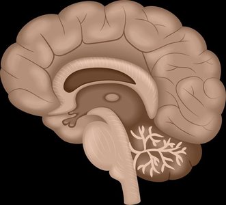
Central Nervous System (CNS): Consists of the brain and spinal cord; integrates and processes information.
Peripheral Nervous System (PNS): Composed of nerves and ganglia outside the CNS; transmits sensory and motor signals.
Main Cell Types in Nervous Tissue
Nervous tissue contains two primary cell types: neurons and glial cells. Each plays a distinct role in the function and maintenance of the nervous system.
Neurons: Excitable cells that transmit electrical signals.
Glial Cells: Support, protect, and nourish neurons; include astrocytes, oligodendrocytes, microglia, and Schwann cells.
Structure of a Neuron
Neurons are specialized for rapid communication. Their structure enables the transmission of electrical impulses.
Cell Body (Soma): Contains the nucleus and metabolic machinery.
Dendrites: Receive incoming signals.
Axon: Conducts impulses away from the cell body.
Axon Terminals: Release neurotransmitters to communicate with other cells.
Classification of Neurons
Neurons are classified based on structure and function. Major connective tissues support and protect these cells.
Structural Classification: Multipolar, bipolar, and unipolar neurons.
Functional Classification: Sensory (afferent), motor (efferent), and interneurons.
Membrane Potential and Ion Channels
Resting Membrane Potential
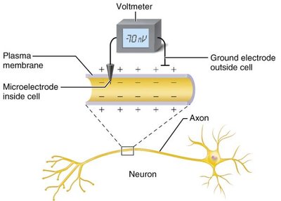
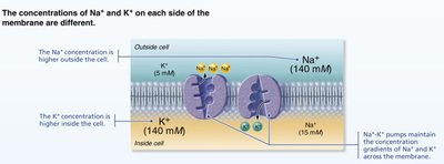
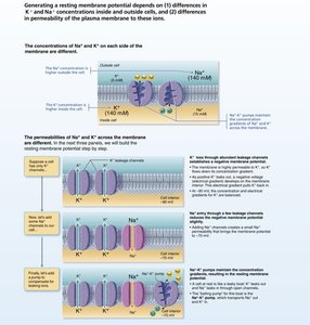
Neurons maintain a resting membrane potential, typically around -70 mV, due to differences in ion concentrations and membrane permeability.
Key ions: Sodium (Na+) and Potassium (K+).
Polarization: The inside of the cell is more negative than the outside.
Generated by: Differences in ionic composition and permeability.
Types of Ion Channels
Ion channels are essential for generating and propagating electrical signals in neurons. They are classified based on their gating mechanisms.
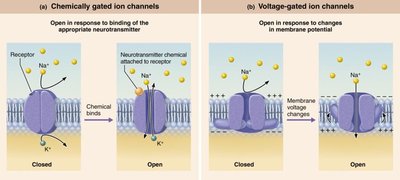
Leakage (nongated) channels: Always open, allow passive ion movement.
Gated channels: Open/close in response to specific stimuli.
Chemically gated (ligand-gated): Open with binding of a neurotransmitter.
Voltage-gated: Open/close in response to changes in membrane potential.
Mechanically gated: Open/close in response to physical deformation.
Graded Potentials and Action Potentials
Graded Potentials
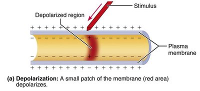
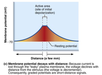
Graded potentials are local changes in membrane potential that occur in response to stimuli. They are essential for initiating action potentials.
Short-distance signals: Occur in dendrites and cell body.
Depolarization: Membrane becomes less negative.
Hyperpolarization: Membrane becomes more negative.
Decay with distance: Voltage declines as current flows away from the site of stimulus.
Action Potentials
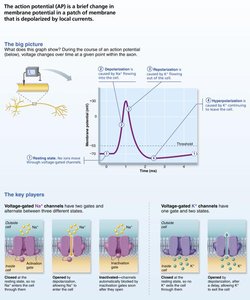
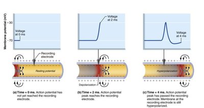
Action potentials are rapid, long-distance electrical signals generated by neurons. They involve a brief reversal of membrane potential and are essential for nerve impulse transmission.
All-or-none phenomenon: Action potential either occurs fully or not at all.
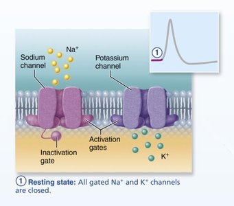
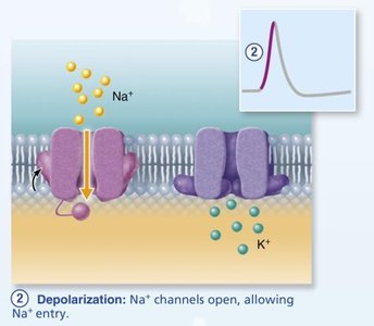
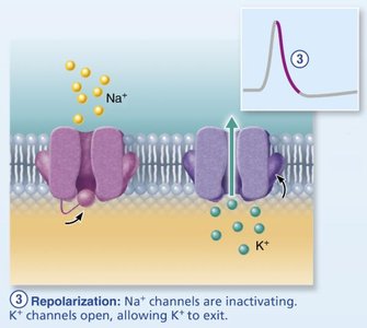
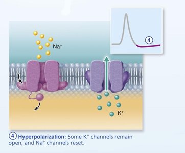
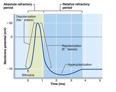
Four main steps: Resting state, depolarization, repolarization, hyperpolarization.
Propagation: Action potential travels along the axon toward terminals.
Refractory Periods
Refractory periods ensure unidirectional propagation and regulate the frequency of action potentials.
Absolute refractory period: No new action potential can be triggered.
Relative refractory period: Only exceptionally strong stimuli can trigger an action potential.
Synapses: Structure and Function
Chemical and Electrical Synapses
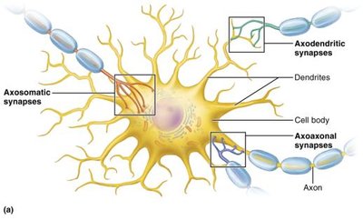
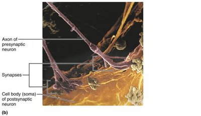
Synapses are junctions where neurons communicate with other neurons or effector cells. They are classified as chemical or electrical based on their mechanism of signal transmission.
Chemical synapses: Use neurotransmitters for signal transmission; unidirectional.
Electrical synapses: Use gap junctions for rapid, bidirectional communication.
Neurotransmitters
Neurotransmitters are chemical messengers released at synapses. They are classified chemically and functionally, and their effects can be excitatory or inhibitory.
Acetylcholine (ACh): Released at neuromuscular junctions; degraded by acetylcholinesterase.
Catecholamines: Dopamine, norepinephrine, epinephrine; involved in stress response.
Indolamines: Serotonin, histamine; regulate mood, sleep, cognition, and other functions.
Clinical Implications
Disorders and drugs affecting nervous tissue and synapses have significant clinical consequences.
Multiple sclerosis: Autoimmune destruction of CNS myelin; impairs impulse conduction.
Local anesthetics: Block voltage-gated Na+ channels, preventing nerve impulses.
Opioid use disorder: Chronic opioid use alters neurotransmitter systems; naloxone can reverse overdose.
Summary Table: Steps of Action Potential
Step | Key Event | Ion Channel Activity |
|---|---|---|
Resting State | Membrane at -70 mV | All gated Na+ and K+ channels closed |
Depolarization | Membrane potential rises to +30 mV | Na+ channels open, Na+ enters cell |
Repolarization | Membrane potential returns toward -70 mV | Na+ channels inactivate, K+ channels open, K+ exits cell |
Hyperpolarization | Membrane potential dips below -70 mV | Some K+ channels remain open, Na+ channels reset |
Key Equations
The resting membrane potential is determined by the Nernst equation:
Where: Eion = equilibrium potential for the ion R = gas constant T = temperature (Kelvin) z = charge of the ion F = Faraday's constant [ion]outside = ion concentration outside the cell [ion]inside = ion concentration inside the cell
Action potential propagation is governed by changes in membrane potential:
Where: ΔV = change in voltage I = current R = resistance
Additional info: Academic context and explanations were expanded for clarity and completeness. Images were included only when directly relevant to the adjacent content.
