 Indietro
Hearing and Equilibrium: Anatomy & Physiology Study Guide
Guida di studio - Note intelligenti
 Appunti personalizzati basati sui tuoi materiali, ampliati con definizioni chiave, esempi e contesto.
Hearing and Equilibrium
Regions and Anatomy of the Ear
The ear is divided into three main regions, each with distinct anatomical features and functions related to hearing and equilibrium.
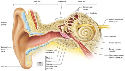
Outer (External) Ear: Includes the auricle (pinna), lobule, and external auditory canal. The auricle funnels sound waves into the external auditory canal, which ends at the tympanic membrane. Ceruminous glands in the canal produce cerumen (earwax) to waterproof and protect the canal and tympanic membrane.
Middle Ear: An air-filled chamber within the temporal bone, lined with mucous membrane. It contains three auditory ossicles (malleus, incus, stapes) connected by synovial joints. The stapes attaches to the oval window, marking the boundary between the middle and inner ear. The pharyngotympanic tube connects the middle ear to the nasopharynx, equalizing air pressure.
Inner (Internal) Ear: Also called the bony labyrinth, it contains the membranous labyrinth lined with endolymph and perilymph. The inner ear is responsible for both hearing and equilibrium, with three main regions: cochlea, vestibule, and semicircular canals.
Substructures and Functions of the Ear
Each substructure of the ear has a specific function and connection to either the vestibular or cochlear portion of the vestibulocochlear nerve (CN VIII).
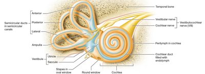
Cochlea: Responsible for hearing; contains the cochlear duct, scala vestibuli, scala tympani, basilar membrane, and spiral organ (organ of Corti). The cochlear branch of CN VIII transmits auditory signals.
Vestibule: Contains utricle and saccule, which detect head position and linear movement. The vestibular branch of CN VIII transmits equilibrium signals.
Semicircular Canals: Three canals (anterior, posterior, lateral) detect rotational movement. Each canal contains an ampulla with crista ampullaris (hair cells for rotational equilibrium).
Principles of Sound
Sound is perceived through the movement of air particles, which create waves that are interpreted by the ear.
Pitch: Refers to the frequency of sound waves, measured in hertz (Hz). Higher frequency = higher pitch.
Frequency: Number of vibrations per second; determines pitch.
Amplitude: Loudness of sound, measured in decibels (dB); determined by the difference between high and low pressure.
Human Hearing Range: 20 Hz to 20,000 Hz; can feel vibrations between 4 Hz and 16 Hz.
Transmission of Sound to the Inner Ear
Sound waves are transmitted through the ear in a series of steps:
Sound waves strike the tympanic membrane, causing movement of auditory ossicles and vibration of the oval window.
Vibration of the oval window produces pressure waves in the perilymph (scala vestibuli) and endolymph (cochlear duct), vibrating the basilar membrane.
High-frequency sounds vibrate the basilar membrane where it is narrow and stiff; low-frequency sounds vibrate where it is wide and flexible.
Processing of Sound in the Inner Ear
The basilar membrane's vibration activates the spiral organ, which contains hair cells responsible for detecting sound.
The basilar membrane moves toward the tectorial membrane, bending stereocilia on hair cells.
Bending opens potassium ion channels, depolarizing the hair cell.
Depolarized hair cells release neurotransmitters, triggering action potentials in the cochlear nerve.
Auditory Pathway
Auditory signals are processed through a specific neural pathway:
Action potentials travel through the cochlear portion of CN VIII to the cochlear nuclei at the medulla-pons junction.
Axons synapse on the superior olivary nucleus in the pons.
Stimuli are sent to the inferior colliculus of the midbrain.
Relayed to the medial geniculate nucleus of the thalamus.
Thalamus stimulates neurons in the primary auditory cortex (temporal lobe).
Types of Hearing Loss
Hearing loss can be classified based on the affected region and mechanism:
Conduction Hearing Loss: Problem in outer or middle ear; prevents sound waves from reaching the inner ear. Causes include earwax buildup, infection, perforated tympanic membrane, or ossicle fusion. Usually temporary.
Sensorineural Hearing Loss:
Sensory: Action potentials cannot be generated in cochlea (damaged hair cells, medications, loud sounds). Treatable with hearing aids or cochlear implants.
Neural: Action potentials fail to propagate through cochlear branch or CNS pathways (strokes, tumors). Not treatable with cochlear implants.
Vestibular Sensation and Equilibrium
Equilibrium is maintained by input from vision, proprioceptors, and the vestibular system. Two types of equilibrium are monitored:
Static Equilibrium: Maintains balance when head and body are not moving but head is tilting. Monitored by utricle and saccule.
Dynamic Equilibrium: Maintains balance during movement. Monitored by semicircular ducts.
Utricle and Saccule: Static Equilibrium/Linear Acceleration
Contain maculae (chambers with receptor walls for head position).
Kinocilium and stereocilia are embedded in the otolithic membrane, which contains otoliths (calcium carbonate crystals).
Utricle responds to vertical tilting; saccule responds to up and down movement.
Semicircular Ducts: Rotational Equilibrium
Three ducts (anterior, posterior, lateral) detect rotation in all planes.
Ampulla at the base contains crista ampullaris (hair cells for rotational movement).
Vestibular Sensation Pathway
Vestibular signals are processed through a neural pathway:
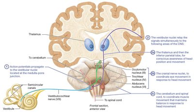
Action potentials propagate to vestibular nuclei at the medulla-pons junction.
Vestibular nuclei relay signals to:
Thalamus and inferior parietal lobe (conscious awareness of head position).
Cranial nerve nuclei (coordinate eye movement).
Cerebellum and spinal cord (coordinate muscle movement for balance).
Clinical Applications
Otitis Media
Inflammation of the middle ear, common in children.
Symptoms: pain, fever, impaired hearing, vomiting.
Causes fluid buildup; tympanic membrane may rupture.
Treatment: antibiotics, myringotomy (surgical drainage).
Tinnitus
Sensation of hearing noise without actual sound input (ringing, buzzing, whistling).
Caused by damage to hair cells, ossicle fusion, tympanic membrane issues, medications, or neural pathway damage.
May be permanent or temporary.
Cochlear Implants
Electrodes bypass damaged hair cells to stimulate cochlear nerve directly.
External microphone and processor convert sound waves to electrical signals.
Signals transmitted to internal receiver and electrodes in cochlea.
Motion Sickness
Caused by mismatched sensory information from eyes and vestibular system.
Symptoms: nausea, dizziness, vomiting.
Treatment: antihistamines, scopolamine patch, reducing conflicting information.
Key Terms and Definitions
Auricle (pinna): External part of the ear that collects sound waves.
Cerumen: Earwax produced by ceruminous glands.
Tympanic membrane: Eardrum; vibrates in response to sound.
Auditory ossicles: Malleus, incus, stapes; transmit sound vibrations.
Oval window: Membrane marking boundary between middle and inner ear.
Bony labyrinth: Rigid, bony outer wall of the inner ear.
Membranous labyrinth: Soft tissue lining inside the bony labyrinth.
Endolymph/Perilymph: Fluids in the inner ear; essential for sound and equilibrium transduction.
Utricle/Saccule: Detect linear acceleration and head position.
Semicircular ducts/canals: Detect rotational movement.
Spiral organ (organ of Corti): Contains hair cells for hearing.
Macula: Chamber with receptor cells for static equilibrium.
Otolithic membrane: Gelatinous layer containing otoliths.
Crista ampullaris: Cluster of hair cells in ampulla for rotational equilibrium.
Summary Table: Ear Regions and Functions
Region | Main Structures | Function |
|---|---|---|
Outer Ear | Auricle, External Auditory Canal, Tympanic Membrane | Collects and funnels sound waves |
Middle Ear | Malleus, Incus, Stapes, Pharyngotympanic Tube | Transmits and amplifies sound; equalizes pressure |
Inner Ear | Cochlea, Vestibule, Semicircular Canals | Hearing (cochlea); Equilibrium (vestibule, semicircular canals) |
Summary Table: Types of Hearing Loss
Type | Location | Causes | Treatment |
|---|---|---|---|
Conduction | Outer/Middle Ear | Earwax, infection, ossicle fusion | Usually temporary |
Sensorineural (Sensory) | Inner Ear | Hair cell damage, medications, loud sounds | Hearing aids, cochlear implants |
Sensorineural (Neural) | Cochlear nerve/CNS | Strokes, tumors | Not treatable with implants |
Key Equations
Frequency (Hz):
Amplitude (dB):
Practice Application
Question: Which structure of the middle ear is the reason when someone has a cold they can experience post nasal drip?
Answer: The pharyngotympanic tube (auditory tube) connects the middle ear to the nasopharynx and can be affected during a cold.
Additional info: Academic context was added to clarify the functions and clinical relevance of each ear region and pathway.
